. 2009 Nov 1;75(3):843-53.
doi: 10.1016/j.ijrobp.2008.06.1953. Epub 2008 Oct 11.

Radiation increases invasion of gene-modified mesenchymal stem cells into tumors

Affiliations

Radiation increases invasion of gene-modified mesenchymal stem cells into tumors

Steven P Zielske et al. Int J Radiat Oncol Biol Phys. .

Abstract

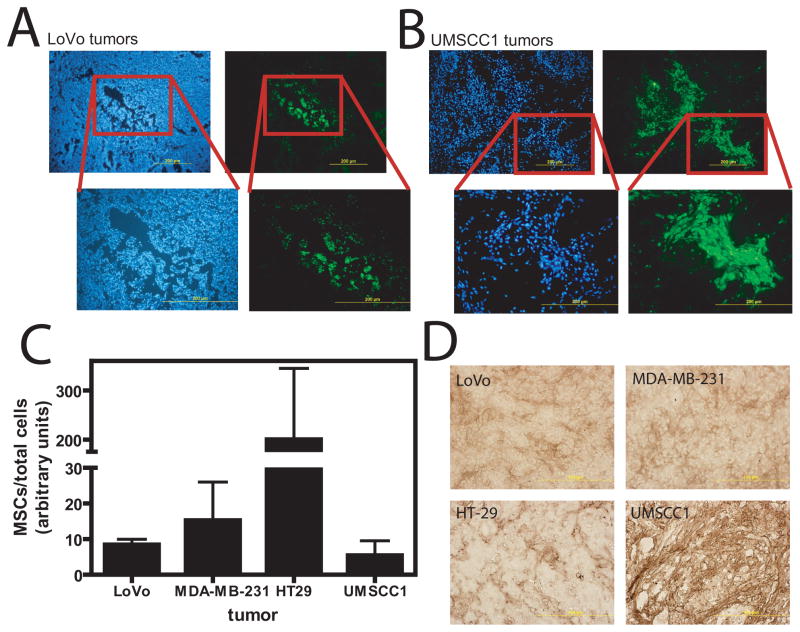
Purpose: Mesenchymal stem cells (MSCs) are multipotent cells in the bone marrow that have been found to migrate to tumors, suggesting a potential use for cancer gene therapy. MSCs migrate to sites of tissue damage, including normal tissues damaged by radiation. In this study, we investigated the effect of tumor radiotherapy on the localization of lentivirus-transduced MSCs to tumors.

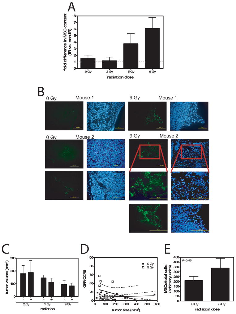
Methods and materials: MSCs were labeled with a lipophilic dye to investigate their migration to colon cancer xenografts. Subsequently, the MSCs were transduced with a lentiviral vector to model gene therapy and mark the infused MSCs. LoVo tumor xenografts were treated with increasing radiation doses to assess the effect on MSC localization, which was measured by quantitative polymerase chain reaction. MSC invasion efficiency was determined in an invasion assay.

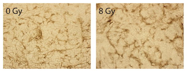
Results: MSCs migrated to tumor xenografts of various origins, with few cells found in normal tissues. A lentiviral vector efficiently transduced MSCs in the presence, but not the absence, of hexadimethrine bromide (Polybrene). When LoVo tumors were treated with increasing radiation doses, more MSCs were found to migrate to them than to untreated tumors. Irradiation increased MSC localization in HT-29 and MDA-MB-231, but not UMSCC1, xenografts. Monocyte chemotactic protein-1 expression in tumors did not correlate with the basal levels of MSC infiltration; however, monocyte chemotactic protein-1 was modestly elevated in irradiated tumors. Media from irradiated LoVo cells stimulated MSC invasion into basement membranes.

Conclusion: These findings suggest that radiation-induced injury can be used to target MSCs to tumors, which might increase the effectiveness of MSC cancer gene therapy. The production of tumor-derived factors in response to radiation stimulates MSC invasion.


Figures

Figure 1
Figure 1
Migration of MSCs to tumors. (A) MSCs were stained with SP-DiOC18 before infusion. LoVo tumor sections from mice receiving labeled MSC infusion 8 d prior. Left column shows 3 different tumor sections with green MSCs located in clusters within the tumor. Right column is the corresponding Dapi picture of the same section showing nuclear staining. It can be seen that clusters are located both internally (top 2 pairs) and at the tumor periphery (bottom pair). (B) Analysis of other tissues for the presence of MSCs shows few to none in liver, spleen, muscle, and bone marrow. Small numbers are observed in the lung at 8 d post-infusion.
Figure 2
Figure 2
MSC transduction. (A) MSCs were transduced with a lentiviral vector in the presence or absence of Polybrene at various MOIs from 2 – 61 and the proportion of GFP+ cells analyzed by flow cytometry. Efficient transduction was only observed in the presence of Polybrene. (B) GFP expression by MSCs in vitro was observed at 4 d post-transduction and maintained for up to 12 wks.
Figure 3
Figure 3
Migration of transduced MSCs to tumors. Lentivirus-transduced MSCs were infused into LoVo or UMSCC1 tumor-bearing mice and tumor sections analyzed at 14 d. (A) Lentivirus-transduced MSCs migrated to, and expressed in, LoVo tumors. (B) UMSCC1 tumors contained several large clusters of GFP+ MSCs. Left panels are Dapi-stained photos of the same section. Right panels are direct GFP fluorescence. Boxed regions define area of higher magnification. (C) Plot of the proportion of MSCs in different tumor types as determined by PCR. A greater proportion of MSCs were found in HT-29 tumors compared to LoVo (P<0.001), MDA-MB-231 (P<0.01), or UMSCC1 (P<0.01; Tukey’s Multiple Comparison test). (D) MCP-1 immunohistochemistry of tumor sections showing different degrees of MCP-1 expression depending on tumor type.
Figure 4
Figure 4
Radiation increases MSC migration to tumors. (A) Radiation dose response of MSCs in LoVo tumors. A greater proportion of MSCs were found in tumors receiving a greater radiation dose (P=0.026 0 Gy vs 9 Gy, t test with Welch’s correction; N=2–11). (B) Photomicrograph examples of tumor sections taken from 0 Gy and 9 Gy irradiated LoVo tumors showing more readily observed MSC in irradiated tumors compared to unirradiated. Left panels are direct GFP fluorescence and right panels are DAPI-stained. (C) Graph of LoVo tumor volume for each irradiated group and their matched unirradated controls. There was no significant difference between groups (P=0.5–0.9, paired t test). (D) Plot of the proportion of MSCs in each tumor versus tumor size. Regression analysis showed no relationship between MSC content and size for neither 0 Gy (P=0.63) nor 9 Gy (P=0.21) irradiated tumors. There was no difference between these 2 groups (P=0.10). (E) MSCs in unirradiated or 8 Gy irradiated skin. MSC content trended higher in irradiated skin, but was not significant (P=0.46).
Figure 5
Figure 5
MCP-1 expression in irradiated tumors. Sections of 0 or 8 Gy irradiated tumors were taken from mice 14 d post-irradiation and stained for MCP-1. In 8 Gy irradiated tumors, a modest increase in MCP-1 staining is observed.
Figure 6
Figure 6
MSCs localize near tumor vasculature. Sections of irradiated tumors taken from mice infused with luciferase-transduced MSCs were co-labeled with anti-CD31 and anti-luciferase antibodies. CD31 and luciferase labeling colocalized, indicating MSCs were predominantly found near tumor vasculature.

References

    1. Zhang M, Li S, Nyati MK, et al. Regional delivery and selective expression of a high-activity yeast cytosine deaminase in an intrahepatic colon cancer model. Cancer Res. 2003;63:658–663. - PubMed
    1. Reid T, Warren R, Kirn D. Intravascular adenoviral agents in cancer patients: lessons from clinical trials. Cancer Gene Ther. 2002;9:979–986. - PMC - PubMed
    1. Herzog EL, Chai L, Krause DS. Plasticity of marrow-derived stem cells. Blood. 2003;102:3483–3493. - PubMed
    1. Pittenger MF, Mackay AM, Beck SC, et al. Multilineage potential of adult human mesenchymal stem cells. Science. 1999;284:143–147. - PubMed
    1. Nakamizo A, Marini F, Amano T, et al. Human bone marrow-derived mesenchymal stem cells in the treatment of gliomas. Cancer Res. 2005;65:3307–3318. - PubMed

Publication types

MeSH terms