. 2008 Dec 5;283(49):33874-82.
doi: 10.1074/jbc.M804016200. Epub 2008 Oct 10.

Na-K-Cl Cotransporter-1 in the mechanism of ammonia-induced astrocyte swelling

Affiliations

Na-K-Cl Cotransporter-1 in the mechanism of ammonia-induced astrocyte swelling

Arumugam R Jayakumar et al. J Biol Chem. .

Abstract

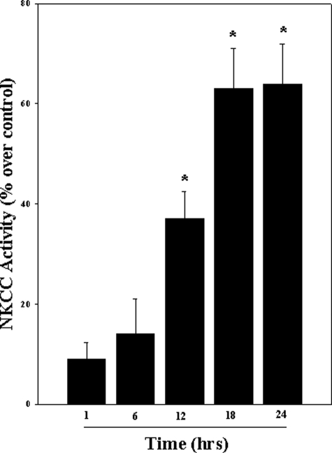
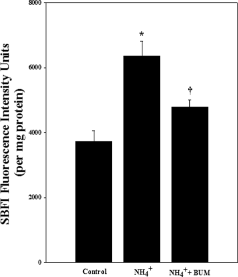
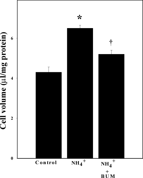
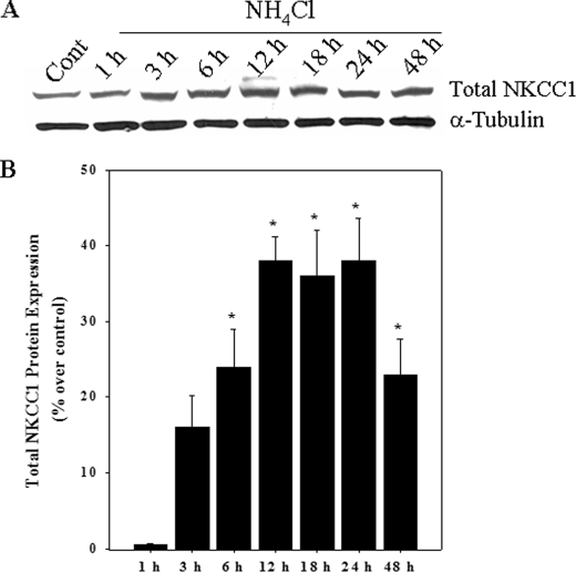
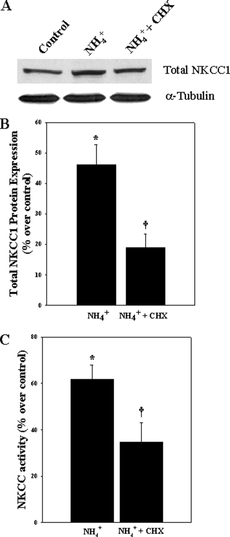
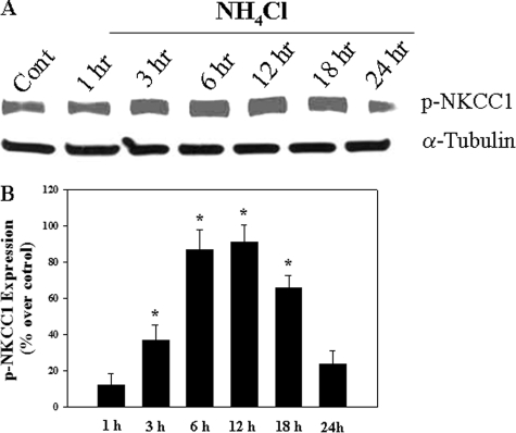
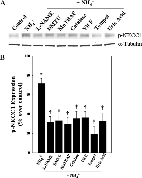
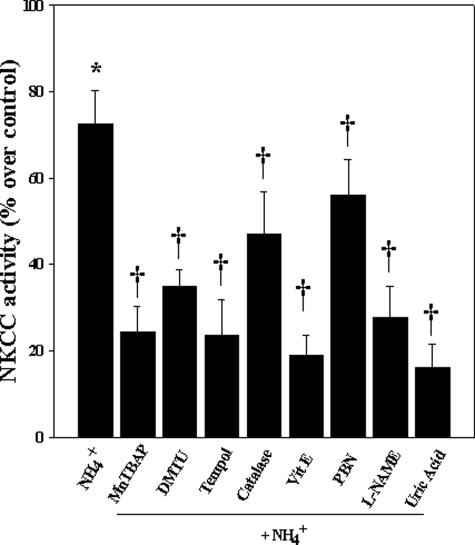
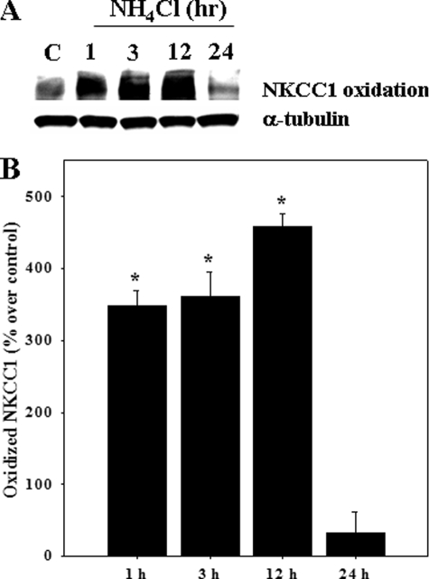
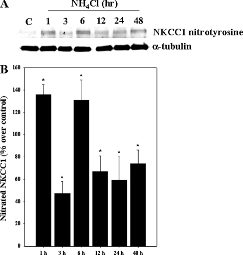
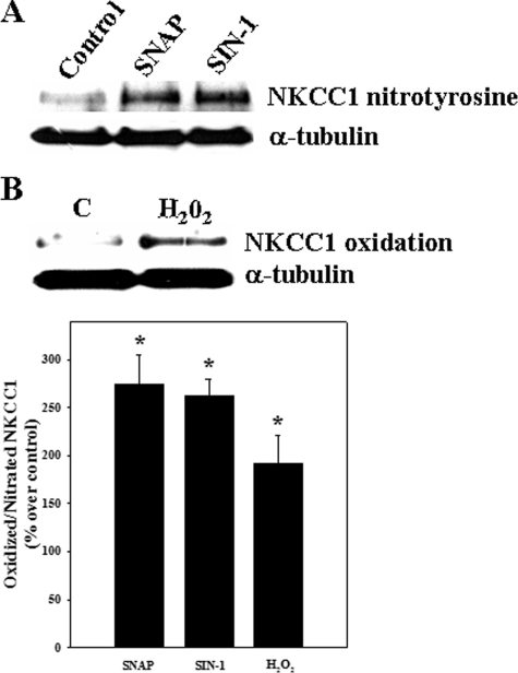
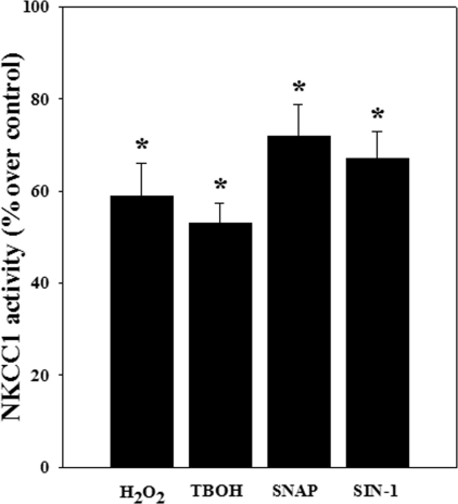
Brain edema and the consequent increase in intracranial pressure and brain herniation are major complications of acute liver failure (fulminant hepatic failure) and a major cause of death in this condition. Ammonia has been strongly implicated as an important factor, and astrocyte swelling appears to be primarily responsible for the edema. Ammonia is known to cause cell swelling in cultured astrocytes, although the means by which this occurs has not been fully elucidated. A disturbance in one or more of these systems may result in loss of ion homeostasis and cell swelling. In particular, activation of the Na-K-Cl cotransporter (NKCC1) has been shown to be involved in cell swelling in several neurological disorders. We therefore examined the effect of ammonia on NKCC activity and its potential role in the swelling of astrocytes. Cultured astrocytes were exposed to ammonia (NH(4)Cl; 5 mm), and NKCC activity was measured. Ammonia increased NKCC activity at 24 h. Inhibition of this activity by bumetanide diminished ammonia-induced astrocyte swelling. Ammonia also increased total as well as phosphorylated NKCC1. Treatment with cyclohexamide, a potent inhibitor of protein synthesis, diminished NKCC1 protein expression and NKCC activity. Since ammonia is known to induce oxidative/nitrosative stress, and antioxidants and nitric-oxide synthase inhibition diminish astrocyte swelling, we also examined whether ammonia caused oxidation and/or nitration of NKCC1. Cultures exposed to ammonia increased the state of oxidation and nitration of NKCC1, whereas the antioxidants N-nitro-l-arginine methyl ester and uric acid all significantly diminished NKCC activity. These agents also reduced phosphorylated NKCC1 expression. These results suggest that activation of NKCC1 is an important factor in the mediation of astrocyte swelling by ammonia and that such activation appears to be mediated by NKCC1 abundance as well as by its oxidation/nitration and phosphorylation.


Figures

FIGURE 1.
FIGURE 1.
Cultured astrocytes were exposed to 5 mm NH4Cl for different time periods (1–24 h), and the bumetanide-sensitive NKCC activity was measured. Ammonia significantly increased NKCC activity at 12, 18, and 24 h. Data were subjected to ANOVA (n = 5; *, p < 0.05 versus control). Error bars, mean ± S.E.
FIGURE 2.
FIGURE 2.
Cultures were exposed to 5 mm NH4Cl for 24 h and [Na+]i was measured by using the Na+ fluorescent probe sodium-binding benzofuran isophthalate. Ammonia significantly increased [Na+]i. Treatment with bumetanide (BUM; 50 μm), an inhibitor of NKCC, significantly diminished this effect. Data were subjected to ANOVA (n = 3; *, p < 0.05 versus control; †, p < 0.05 versus NH4Cl). Error bars, mean ± S.E.
FIGURE 3.
FIGURE 3.
Cultured astrocytes exposed to 5 mm NH4Cl significantly increased cell swelling (54%) at 24 h. Treatment with the NKCC inhibitor bumetanide (BUM; 50 μm) significantly diminished such swelling (65%). Data were subjected to ANOVA (n = 6; *, p < 0.05 versus control; †, p < 0.05 versus NH4Cl). Error bars, mean ± S.E.
FIGURE 4.
FIGURE 4.
A, Western blots show a significant increase in total NKCC1 protein level when cultures were exposed to 5 mm NH4Cl. B, quantification of NH4Cl-induced changes in NKCC1 protein expression. NKCC1 levels were normalized against α-tubulin. Data were subjected to ANOVA (n = 5; *, p < 0.05 versus control). Error bars, mean ± S.E.
FIGURE 5.
FIGURE 5.
A, Western blots show the effect of CHX on ammonia-induced total NKCC1 protein level. B, quantification of NKCC1 protein expression. NKCC1 levels were normalized against α-tubulin. C, effect of cyclohexamide on ammonia-induced NKCC activity. Data were subjected to ANOVA (n = 5; *, p < 0.05 versus control; †, p < 0.05 versus NH4Cl). Error bars, mean ± S.E.
FIGURE 6.
FIGURE 6.
A, Western blots show a significant increase in p-NKCC1 level when cultures were exposed to 5 mm NH4Cl. B, quantification of NH4Cl-induced phospho-NKCC1. Phospho-NKCC1 levels were normalized against α-tubulin. Data were subjected to ANOVA (n = 4; *, p < 0.05 versus control). Error bars, mean ± S.E.
FIGURE 7.
FIGURE 7.
Effect of antioxidants, l-NAME, and uric acid on p-NKCC1 expression. Astrocytes were pretreated (15 min) with dimethylthiourea (DMTU; 100 μm), Mn(III) tetrakis (4-benzoic acid) porphyrin (MnTBAP; 10 μm), catalase (250 units/ml), α-tocopherol (100 μm), tempol (10 μm), l-NAME (250 μm), and uric acid (500 μm) and exposed to ammonia for 24 h, and p-NKCC1 levels were measured. Antioxidants, l-NAME, and uric acid significantly inhibited p-NKCC1 expression. Data were subjected to ANOVA (n = 4; *, p < 0.05 versus control; †, p < 0.05 versus NH4Cl). Error bars, mean ± S.E.
FIGURE 8.
FIGURE 8.
Effect of antioxidants, l-NAME, and uric acid on NKCC1 activity. Astrocytes were treated with Mn(III) tetrakis (4-benzoic acid) porphyrin (MnT-BAP; 10 μm), dimethylthiourea (DMTU; 100 μm), tempol (10 μm), catalase (250 units/ml), α-tocopherol (100 μm), l-NAME (250 μm), and uric acid (500 μm) for 24 h, and NKCC1 activity was measured. Antioxidants, l-NAME, and uric acid significantly inhibited NKCC1 activity. Data were subjected to ANOVA (n = 5; *, p < 0.05 versus control; †, p < 0.05 versus NH4Cl). Error bars, mean ± S.E.
FIGURE 9.
FIGURE 9.
Cultured astrocytes were treated with 5 mm NH4Cl for different time periods (1–24 h), and oxidized NKCC1 protein was determined by using “OxyBlot.” A, Western blots show a significant increase in oxidized NKCC1 at 1, 3, and 12 h after ammonia treatment. Lane C, control. B, quantification of ammonia-induced NKCC1 oxidative adduct formation. Oxidized NKCC1 levels are normalized against α-tubulin. Data were subjected to ANOVA (n = 4; *, p < 0.05 versus control). Error bars, mean ± S.E.
FIGURE 10.
FIGURE 10.
Cultured astrocytes were treated with 5 mm NH4Cl for different time periods (1–48 h), and nitrated NKCC1 was measured. A, Western blots show a significant increase in nitrated NKCC1 from 1–48 h after ammonia treatment. Lane C, control. B, quantification of ammonia-induced NKCC1 nitration. Nitrated NKCC1 levels are normalized against α-tubulin. Data were subjected to ANOVA (n = 5; *, p < 0.05 versus control). Error bars, mean ± S.E.
FIGURE 11.
FIGURE 11.
Astrocyte cultures were treated with hydrogen peroxide (H2O2; 250 μm) and nitric oxide donors (SNAP (200 μm) and SIN-1 (500 μm)), and nitrated NKCC1 was measured at 3 h. A, both SNAP and SIN-1 increased protein tyrosine nitration. B, H2O2 increased oxidized NKCC1. C, quantification of ammonia-induced NKCC1 oxidation/nitration. Nitrated NKCC1 levels are normalized against α-tubulin. Data were subjected to ANOVA (n = 5; *, p < 0.05 versus control). Error bars, mean ± S.E.
FIGURE 12.
FIGURE 12.
NKCC1 activity after exposure of astrocyte cultures to oxidants and NO donors. Astrocytes were treated with H2O2 (25 μm), tert-butylhydroperoxide (TBOH) (25 μm), SNAP (50 μm), and SIN-1 (100 μm) for 24 h, and NKCC1 activity was measured. Both oxidants (H2O2 (58%) and tert-butylhydroperoxide (53%)) and NO donors (SNAP (71%) and SIN-1 (64%)) significantly increased NKCC1 activity. Data were subjected to ANOVA (n = 4; *, p < 0.05 versus control). Error bars, mean ± S.E.

References

    1. Vaquero, J., Chung, C., and Blei, A. T. (2003) Ann. Hepatol. 2 12–22 - PubMed
    1. Albrecht, J., and Jones, E. A. (1999) J. Neurol. Sci. 170 138–146 - PubMed
    1. Hazell, A. S., and Butterworth, R. F. (1999) Proc. Soc. Exp. Biol. Med. 222 99–112 - PubMed
    1. Martinez, A. J. (1968) Acta Neuropathol. (Berl.) 11 82–86 - PubMed
    1. Norenberg, M. D. (1977) Lab. Invest. 36 618–627 - PubMed

Publication types

MeSH terms

LinkOut - more resources