. 2009 Mar;50(3):521-533.
doi: 10.1194/jlr.M800388-JLR200. Epub 2008 Oct 10.

A novel role for activating transcription factor-2 in 15(S)-hydroxyeicosatetraenoic acid-induced angiogenesis

Affiliations

A novel role for activating transcription factor-2 in 15(S)-hydroxyeicosatetraenoic acid-induced angiogenesis

Tieqiang Zhao et al. J Lipid Res. 2009 Mar.

Abstract

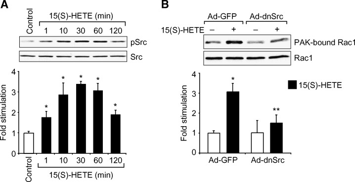
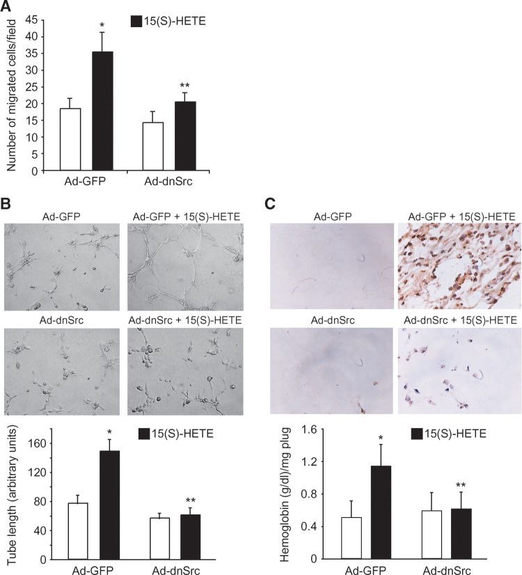
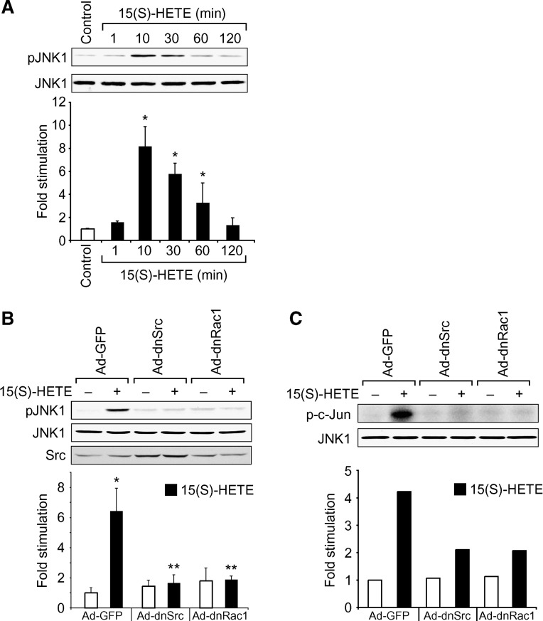
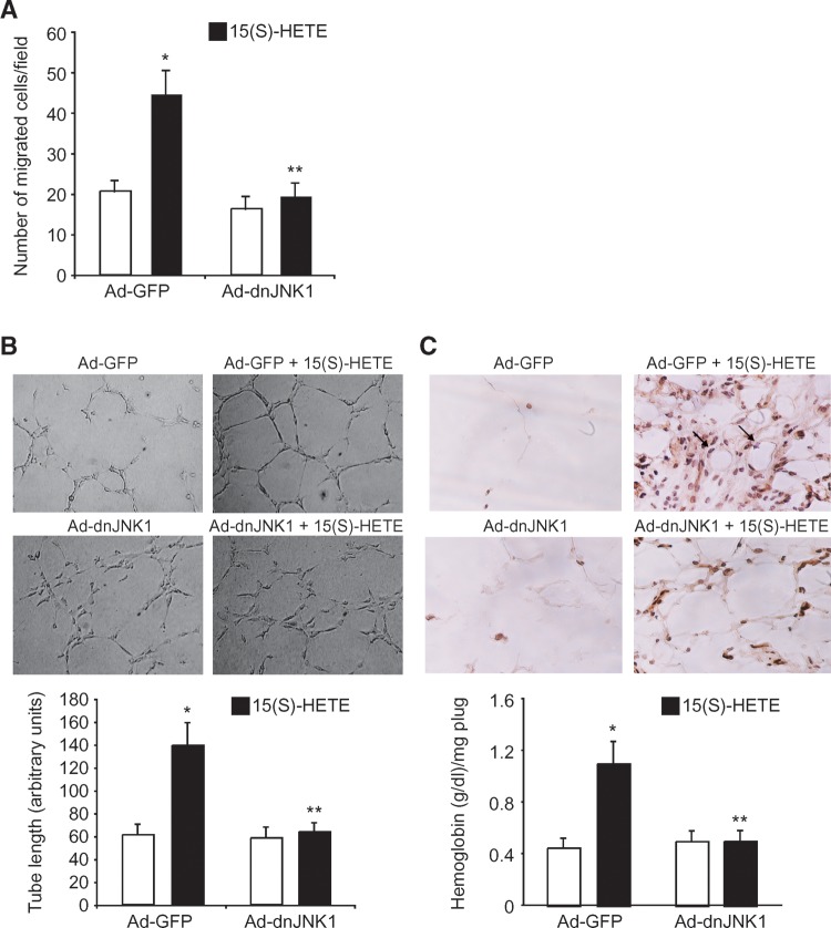
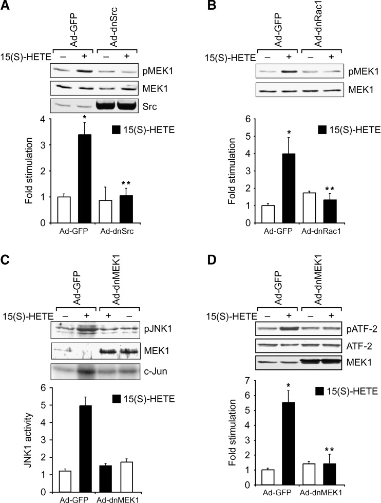
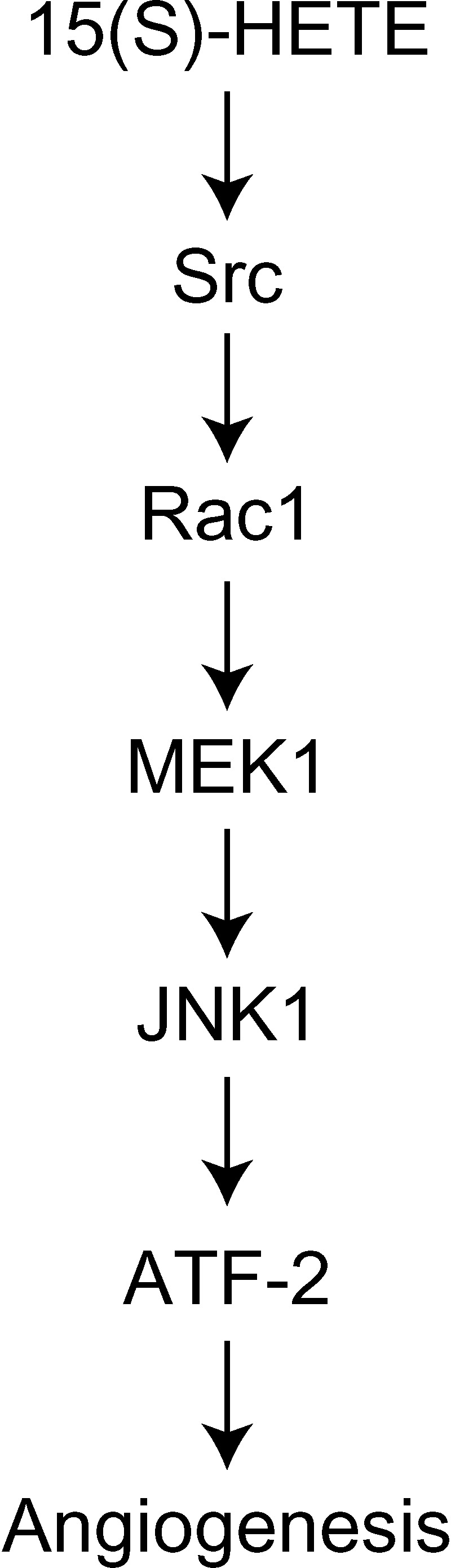
To investigate the mechanisms underlying 15(S)-HETE-induced angiogenesis, we have studied the role of the small GTPase, Rac1. We find that 15(S)-HETE activated Rac1 in human retinal microvascular endothelial cells (HRMVEC) in a time-dependent manner. Blockade of Rac1 by adenovirus-mediated expression of its dominant negative mutant suppressed HRMVEC migration as well as tube formation and Matrigel plug angiogenesis. 15(S)-HETE stimulated Src in HRMVEC in a time-dependent manner and blockade of its activation inhibited 15(S)-HETE-induced Rac1 stimulation in HRMVEC and the migration and tube formation of these cells as well as Matrigel plug angiogenesis. 15(S)-HETE stimulated JNK1 in Src-Rac1-dependent manner in HRMVEC and adenovirus-mediated expression of its dominant negative mutant suppressed the migration and tube formation of these cells and Matrigel plug angiogenesis. 15(S)-HETE activated ATF-2 in HRMVEC in Src-Rac1-JNK1-dependent manner and interference with its activation via adenovirus-mediated expression of its dominant negative mutant abrogated migration and tube formation of HRMVEC and Matrigel plug angiogenesis. In addition, 15(S)-HETE-induced MEK1 stimulation was found to be dependent on Src-Rac1 activation. Blockade of MEK1 activation inhibited 15(S)-HETE-induced JNK1 activity and ATF-2 phosphorylation. Together, these findings show that 15(S)-HETE activates ATF-2 via the Src-Rac1-MEK1-JNK1 signaling axis in HRMVEC leading to their angiogenic differentiation.


Figures

Fig. 1.
Fig. 1.
15(S)-HETE-induced angiogenic differentiation of human retinal microvascular endothelial cells (HRMVEC) requires Rac1 activation. A: Quiescent HRMVEC were treated with and without 15(S)-HETE (0.1 μM) for the indicated times and cell extracts were prepared and analyzed for Rac1 activation by pull-down assay. Cellular total Rac1 levels are shown in the lower panel. B, C: HRMVEC were transduced with Ad-GFP or Ad-dnRac1 at a moi of 80, quiesced, and subjected to 15(S)-HETE-induced migration (B) or tube formation (C). D: C57BL/6 mice were injected subcutaneously with 0.5 ml of Matrigel premixed with vehicle or 50 μM 15(S)-HETE with and without Ad-GFP or Ad-dnRac1 (5 × 10⁹ pfu/ml). One week later, the animals were sacrificed, and the Matrigel plugs were harvested from underneath the skin and either immunostained for CD31 expression using anti-CD31 antibodies or analyzed for hemoglobin content using Drabkin's reagent. The values in the bar graphs in panels A, B, C, and D are the means ± SD of three independent experiments or four plugs from four animals. * P < 0.01 vs. control or Ad-GFP; ** P < 0.01 vs. Ad-GFP + 15(S)-HETE. The white bars represent controls to their respective treatments.
Fig. 2.
Fig. 2.
Src mediates 15(S)-HETE-induced Rac1 activation in HRMVEC. A: Quiescent HRMVEC were treated with and without 15(S)-HETE (0.1 μM) for the indicated times and cell extracts were prepared and analyzed for Src activation by Western blotting using its phospho-specific antibodies. For normalization, the same blot was reprobed with anti-Src antibodies. B: HRMVEC were transduced with Ad-GFP or Ad-dnSrc at a moi of 80, quiesced, treated with and without 15(S)-HETE (0.1 μM) for 10 min, and cell extracts were prepared and analyzed for Rac1 activation by pull-down assay. The values in the bar graphs in panels A and B are the means ± SD of three independent experiments. * P < 0.01 vs. control or Ad-GFP; ** P < 0.01 vs. Ad-GFP + 15(S)-HETE. The white bars represent controls to their respective treatments.
Fig. 3.
Fig. 3.
Blockade of Src activation suppresses 15(S)-HETE-induced HRMVEC migration and tube formation in vitro and Matrigel plug angiogenesis in vivo. A, B: HRMVEC were transduced with Ad-GFP or Ad-dnSrc at a moi of 80, quiesced, and subjected to 15(S)-HETE-induced migration (A) or tube formation (B). C: C57BL/6 mice were injected subcutaneously with 0.5 ml of Matrigel premixed with vehicle or 50 μM 15(S)-HETE with and without Ad-GFP or Ad-dnSrc (5 × 10⁹ pfu/ml). One week later, the animals were sacrificed, and the Matrigel plugs were harvested from underneath the skin and either immunostained for CD31 expression using anti-CD31 antibodies or analyzed for hemoglobin content using Drabkin's reagent. The values in the bar graphs in panels A, B, and C are the means ± SD of three independent experiments or four plugs from four animals. * P < 0.01 vs. Ad-GFP; ** P < 0.01 vs. Ad-GFP + 15(S)-HETE. The white bars represent controls to their respective treatments.
Fig. 4.
Fig. 4.
Src-Rac1 signaling mediates 15(S)-HETE-induced JNK1 activation in HRMVEC. A: Quiescent HRMVEC were treated with and without 15(S)-HETE (0.1 μM) for the indicated times, and cell extracts were prepared and analyzed by Western blotting for pJNK1 using its phosphospecific antibodies. The blot was reprobed with anti-JNK1 antibodies for normalization. B, C: HRMVEC were transduced with Ad-GFP, Ad-dnSrc, or Ad-dnRac1 at a moi of 80, quiesced, treated with and without 15(S)-HETE (0.1 μM) for 10 min, and cell extracts were prepared and analyzed for JNK1 activation either by Western blotting using its phosphospecific antibodies or by immunocomplex kinase assay using GST-c-Jun protein and [γ-32P]ATP as substrates. The blots in panels A and B were reprobed either with anti-JNK1 antibodies for normalization or anti-Src antibodies to show overexpression of Src. The values in the bar graphs in panels A and B are the means ± SD of three independent experiments. * P < 0.01 vs. control or Ad-GFP; ** P < 0.01 vs. Ad-GFP + 15(S)-HETE. The white bars represent controls to their respective treatments.
Fig. 5.
Fig. 5.
Adenovirus-mediated expression of dnJNK1 inhibits 15(S)-HETE-induced HRMVEC migration and tube formation in vitro and Matrigel plug angiogenesis in vivo. A, B: HRMVEC were transduced with Ad-GFP or Ad-dnJNK1 at a moi of 80, quiesced, and subjected to 15(S)-HETE-induced migration (A) or tube formation (B). C: C57BL/6 mice were injected subcutaneously with 0.5 ml of Matrigel premixed with vehicle or 50 μM 15(S)-HETE with and without Ad-GFP or Ad-dnJNK1 (5 × 10⁹ pfu/ml). One week later, the animals were sacrificed, and the Matrigel plugs were harvested from underneath the skin and either immunostained for CD31 expression using anti-CD31 antibodies or analyzed for hemoglobin content using Drabkin's reagent. The values in the bar graphs in panels A, B, and C are the means ± SD of three independent experiments or four plugs from four animals. * P < 0.01 vs. Ad-GFP; ** P < 0.01 vs. Ad-GFP + 15(S)-HETE. The white bars represent controls to their respective treatments.
Fig. 6.
Fig. 6.
Src-Rac1-JNK1 signaling mediates 15(S)-HETE-induced ATF-2 activation in HRMVEC. A: Quiescent HRMVEC were treated with and without 15(S)-HETE (0.1 μM) for the indicated times, and cell extracts were prepared and analyzed by Western blotting for pATF-2 using its phosphospecific antibodies. B, C: HRMVEC were transduced with Ad-GFP, Ad-dnSrc, Ad-dnRac1, or Ad-dnJNK1 at a moi of 80, quiesced, treated with and without 15(S)-HETE (0.1 μM) for 10 min, and cell extracts were prepared and analyzed for ATF-2 phosphorylation as described in panel A. The blots in panels A, B, and C were reprobed either with anti-ATF-2 antibodies for normalization or anti-Src antibodies to show overexpression of Src. The values in the bar graphs are the means ± SD of three independent experiments. * P < 0.01 vs. control or Ad-GFP; ** P < 0.01 vs. Ad-GFP + 15(S)-HETE. The white bars represent controls to their respective treatments.
Fig. 7.
Fig. 7.
Adenovirus-mediated expression of dnATF-2 suppresses 15(S)-HETE-induced HRMVEC migration and tube formation in vitro and Matrigel plug angiogenesis in vivo. A, B: HRMVEC were transduced with Ad-GFP or Ad-dnATF-2 at a moi of 80, quiesced, and subjected to 15(S)-HETE-induced migration (A) or tube formation (B). C: C57BL/6 mice were injected subcutaneously with 0.5 ml of Matrigel premixed with vehicle or 50 μM 15(S)-HETE with and without Ad-GFP or Ad-dnATF-2 (5 × 10⁹ pfu/ml). One week later, the animals were sacrificed, and the Matrigel plugs were harvested from underneath the skin and either immunostained for CD31 expression using anti-CD31 antibodies or analyzed for hemoglobin content using Drabkin's reagent. The values in the bar graphs in panels A, B, and C are the means ± SD of three independent experiments or four plugs from four animals. * P < 0.01 vs. Ad-GFP; ** P < 0.01 vs. Ad-GFP + 15(S)-HETE. The white bars represent controls to their respective treatments.
Fig. 8.
Fig. 8.
Src-Rac1 signaling targets MEK1 in mediating JNK1-dependent ATF-2 activation in HRMVEC in response to 15(S)-HETE. A, B: HRMVEC were transduced with Ad-GFP, Ad-dnSrc, or Ad-dnRac1 at a moi of 80, quiesced, treated with and without 15(S)-HETE (0.1 μM) for 10 min, and cell extracts were prepared and analyzed for MEK1 activation by Western blotting using its phosphospecific antibodies. The blots were reprobed with anti-MEK1 antibodies for normalization. C, D: HRMVEC were transduced with Ad-GFP or Ad-dnMEK1 at a moi of 80, quiesced, treated with and without 15(S)-HETE (0.1 μM) for 10 min, and cell extracts were prepared and analyzed for JNK1 and ATF-2 phosphorylation using their phospho-specific antibodies. In panel C, an equal amount of protein from control and each treatment was also analyzed for JNK1 activity using immunocomplex kinase assay. In panel D, the blot was reprobed with anti-ATF-2 antibodies for normalization. The blots in panels A, B, C, and D were reprobed either with anti-MEK1 antibodies, anti-Src antibodies, or anti-ATF-2 antibodies for normalization or to show adenovirus-mediated overexpression of Src or MEK1. The values in the bar graphs in panels A, B, C, and D are the means ± SD of three independent experiments. * P < 0.01 vs. Ad-GFP; ** P < 0.01 vs. Ad-GFP + 15(S)-HETE. The white bars represent controls to their respective treatments.
Fig. 9.
Fig. 9.
AA stimulates phosphorylation of ATF-2 in HRMVEC in vitro and induces Matrigel plug angiogenesis in vivo. A: Quiescent HRMVEC were treated with and without AA (5 μM) for the indicated times, and cell extracts were prepared and analyzed by Western blotting for pATF-2 using its phosphospecific antibodies. B, C: C57BL/6 mice were injected subcutaneously with 0.5 ml of Matrigel premixed with vehicle or 50 μM AA. One week later, the animals were sacrificed, and the Matrigel plugs were harvested from underneath the skin and either immunostained for CD31 expression using anti-CD31 antibodies or analyzed for hemoglobin content using Drabkin's reagent. The values in the bar graph are the means ± SD of four plugs from four animals. * P < 0.01 vs. control.
Fig. 10.
Fig. 10.
Schematic diagram showing the potential mechanism of ATF-2 activation by 15(S)-HETE in the stimulation of HRMVEC angiogenic differentiation.

References

    1. Sigal E., C. S. Craik, E. Highland, D. Grunberger, L. L. Costello, R. A. Dixon, and J. A. Nadel. 1988. Molecular cloning and primary structure of human 15-lipoxygenase. Biochem. Biophys. Res. Commun. 157 457–464. - PubMed
    1. Brash A. R., W. E. Boeglin, and M. S. Chang. 1997. Discovery of a second 15(S)-lipoxygenase in humans. Proc. Natl. Acad. Sci. USA. 94 6148–6152. - PMC - PubMed
    1. Bryant R. W., J. M. Bailey, T. Schewe, and S. M. Rapoport. 1982. Positional specificity of a reticulocyte lipoxygenase. Conversion of arachidonic acid to 15(S)-hydroperoxyeicosatetraenoic acid. J. Biol. Chem. 257 6050–6055. - PubMed
    1. Kilty L., A. Logan, and P. J. Vickers. 1999. Differential characteristics of human 15-lipoxygenase isozymes and a novel splice variant of 15S-lipoxygenase. Eur. J. Biochem. 266 83–93. - PubMed
    1. Chang M. S., C. Schneider, R. L. Roberts, S. B. Shappell, F. R. Haselton, W. E. Boeglin, and A. R. Brash. 2005. Detection and subcellular localization of two 15S-lipoxygenases in human cornea. Invest. Ophthalmol. Vis. Sci. 46 849–856. - PubMed

Publication types

MeSH terms