. 2009 Jan 1;113(1):58-65.
doi: 10.1182/blood-2008-07-168393. Epub 2008 Oct 10.

Induction of HIV-1 latency and reactivation in primary memory CD4+ T cells

Affiliations

Induction of HIV-1 latency and reactivation in primary memory CD4+ T cells

Alberto Bosque et al. Blood. .

Abstract

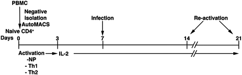
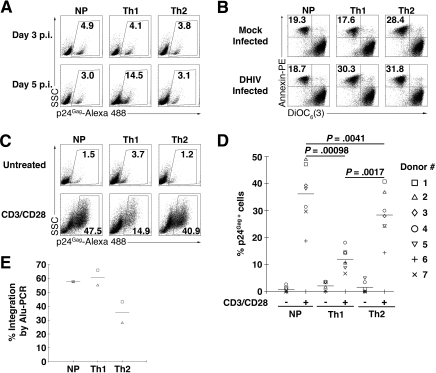
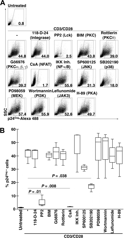
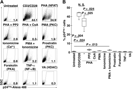
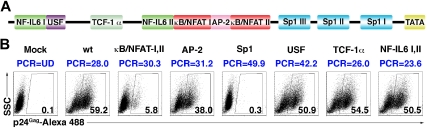
The use of antiretroviral therapy in HIV type 1 (HIV-1)-infected patients does not lead to virus eradication. This is due, to a significant degree, to the fact that HIV-1 can establish a highly stable reservoir of latently infected cells. In this work, we describe an ex vivo experimental system that generates high levels of HIV-1 latently infected memory cells using primary CD4+ T cells. Using this model, we were able to dissect the T cell-signaling pathways and to characterize the long terminal repeat (LTR) cis-acting elements involved in reactivation of HIV-1 in memory CD4+ T cells. We conclude that Lck and nuclear factor of activated T cells (NFAT), but not NF-kappaB, are required for optimal latent virus reactivation in memory T cells. We also found that the cis-acting elements which are critical toward HIV-1 reactivation are the Sp1 and kappaB/NFAT transcription factor binding sites.


Figures

Figure 1
Figure 1
Model of HIV-1 latency. Procedure used for the generation of human primary memory T cells and subsequent establishment of latent infections.
Figure 2
Figure 2
Generation of latently HIV-1–infected primary CD4+ T cells ex vivo. Cells were primed in NP, Th1-, or Th2-polarizing conditions and 7 days after activation cells were infected with DHIV. (A) At 3 and 5 days after infection, cells were assessed for intracellular p24 gag expression by flow cytometry. The percentage of p24-postive cells is indicated in each panel. The experiment shown is representative of 4 different experiments with 4 different donors. (B) At 5 days after infection, cells were assessed for annexin-PE and DiOC6(3) by flow cytometry. For each panel, the percentage of apoptotic cells (annexin-PE positive and DiOC6(3) low) is indicated. The experiment shown is representative of 3 different experiments with 3 different donors. (C) At 7 days after infection, cells were cultured without stimulation (untreated) or costimulated with antibodies to CD3 and CD28 for 3 days (CD3/CD28) and assessed for intracellular p24 gag expression by flow cytometry. The percentage of p24-postive cells is indicated in each panel for this representative experiment. Values corresponding to 7 different donors are shown in panel D, where each symbol represents a different donor and horizontal lines indicate media values. Significance by 2-tailed paired-samples t test analysis (P values provided). (E) Viral integration was analyzed by Alu-PCR 3 days after infection in donors 1 and 2. Horizontal lines indicate media values.
Figure 3
Figure 3
Signaling antagonists and their effects on HIV-1 reactivation. NP cells were infected with DHIV and 7 days after infection cells were left untreated or costimulated with antibodies to CD3 and CD28 for 3 days (CD3/CD28) in the presence of the indicated inhibitor for the protein or pathway indicated between parentheses and assessed for intracellular p24 gag expression by flow cytometry. (A) Representative experiment. The percentage of p24-postive cells is indicated in each panel. (B) Box-plots corresponding to 3 different donors. Horizontal lines indicate median values and significance by 2-tailed paired-samples t test analysis (P values provided).
Figure 4
Figure 4
Signaling agonists and their effects on HIV-1 reactivation. NP cells were infected with DHIV and 7 days after infection cells were left untreated, costimulated (CD3/CD28) in the presence of the indicated agonist for the protein or pathway indicated between parentheses for 3 days, and assessed for intracellular p24 gag expression by flow cytometry. In the case of cells stimulated with PHA, cells were also costimulated in the presence of the inhibitors PP2 (Lck) or CsA (NFAT). (A) Representative experiment. The percentage of p24-postive cells is indicated in each panel. (B) Box-plots corresponding to 3 different donors. Horizontal lines indicate median values and significance by 2-tailed paired-samples t test analysis (P values provided).
Figure 5
Figure 5
Transcription factor binding sites involved in HIV-1 reactivation. (A) Scheme of HIV-1 LTR. (B) NP cells were infected with wt DHIV or with different LTR mutants. Mutations can be viewed in Figure S2. At 7 days after infection, cells were costimulated with antibodies to CD3 and CD28 for 3 days and assessed for intracellular p24 gag expression by flow cytometry. The percentage of p24-postive cells is indicated in each panel. Percentage of viral integration by Alu-PCR for each virus is indicated in blue (UD indicates undetectable). The experiment is representative of 3 different experiments with 3 different donors.

References

    1. Finzi D, Hermankova M, Pierson T, et al. Identification of a reservoir for HIV-1 in patients on highly active antiretroviral therapy. Science. 1997;278:1295–1300. - PubMed
    1. Chun TW, Carruth L, Finzi D, et al. Quantification of latent tissue reservoirs and total body viral load in HIV-1 infection. Nature. 1997;387:183–188. - PubMed
    1. Folks TM, Clouse KA, Justement J, et al. Tumor necrosis factor alpha induces expression of human immunodeficiency virus in a chronically infected T-cell clone. Proc Natl Acad Sci U S A. 1989;86:2365–2368. - PMC - PubMed
    1. Antoni BA, Rabson AB, Kinter A, Bodkin M, Poli G. NF-kappa B-dependent and -independent pathways of HIV activation in a chronically infected T cell line. Virology. 1994;202:684–694. - PubMed
    1. Jordan A, Bisgrove D, Verdin E. HIV reproducibly establishes a latent infection after acute infection of T cells in vitro. Embo J. 2003;22:1868–1877. - PMC - PubMed

Publication types

MeSH terms