. 2008 Oct;20(10):2586-602.
doi: 10.1105/tpc.108.062760. Epub 2008 Oct 10.

Histone H2B monoubiquitination in the chromatin of FLOWERING LOCUS C regulates flowering time in Arabidopsis

Affiliations

Histone H2B monoubiquitination in the chromatin of FLOWERING LOCUS C regulates flowering time in Arabidopsis

Ying Cao et al. Plant Cell. 2008 Oct.

Abstract

Ubiquitination is one of many known histone modifications that regulate gene expression. Here, we examine the Arabidopsis thaliana homologs of the yeast E2 and E3 enzymes responsible for H2B monoubiquitination (H2Bub1). Arabidopsis has two E3 homologs (HISTONE MONOUBIQUITINATION1 [HUB1] and HUB2) and three E2 homologs (UBIQUITIN CARRIER PROTEIN [UBC1] to UBC3). hub1 and hub2 mutants show the loss of H2Bub1 and early flowering. By contrast, single ubc1, ubc2, or ubc3 mutants show no flowering defect; only ubc1 ubc2 double mutants, and not double mutants with ubc3, show early flowering and H2Bub1 defects. This suggests that ubc1 and ubc2 are redundant, but ubc3 is not involved in flowering time regulation. Protein interaction analysis showed that HUB1 and HUB2 interact with each other and with UBC1 and UBC2, as well as self-associating. The expression of FLOWERING LOCUS C (FLC) and its homologs was repressed in hub1, hub2, and ubc1 ubc2 mutant plants. Association of H2Bub1 with the chromatin of FLC clade genes depended on UBC1,2 and HUB1,2, as did the dynamics of methylated histones H3K4me3 and H3K36me2. The monoubiquitination of H2B via UBC1,2 and HUB1,2 represents a novel form of histone modification that is involved in flowering time regulation.


Figures

Figure 1.
Figure 1.
Mutation of HUB1 or HUB2 Leads to an Early Flowering Phenotype in Arabidopsis. (A) Gene structures and characterization of the steady state mRNA levels of HUB1 and HUB2 by RT-PCR in the hub1 and hub2 mutants. Exons are represented by filled black boxes, introns by lines, untranslated regions by filled gray boxes, and T-DNA insertions by triangles. The primers used to detect the transcripts are indicated as P1 to P8. Pairs of P3/P4 and P7/P8 were used to detect the full-length transcripts of HUB1 and HUB2, respectively; Pairs of P1/P2 and P5/P6 were used to detect the partial transcripts of HUB1 and HUB2, respectively. (B) and (C)Phenotypes of hub1 and hub2 single mutants and the hub1 hub2 double mutant at 26 (B) and 42 d old (C). (D) and (E) Complementation of hub1 and hub2 by wild-type (D) (HUB1/hub1 and HUB2/hub2) or mutant (E)(HUB1M/hub1 and HUB2M/hub2) HUB1 or HUB2, respectively. Thirteen out of 15 and 18 out of 20 single copy insertion lines were rescued by the transformation of HUB1 into hub1-5 and hub1-4, respectively, whereas 16 out of 19 single copy insertion lines were rescued by the transformation of HUB2 into hub2-2. No line out of 54 lines for each genotype was rescued by the transformation of HUB1M into hub1-5 and hub1-4, and no line out of 64 lines was rescued by the transformation of HUB2M into hub2-2. Bars = 1 cm.
Figure 2.
Figure 2.
Self-Association and Pairwise Interaction of HUB1 and HUB2. (A) Yeast two-hybrid assay. Positive control: pGADT7-T + pGBKT7-53 (encoding the fusions between GAL4 DNA-BD and AD and murine p53 and SV40 large T-antigen, respectively). p53 and large T-antigen interact in a yeast two-hybrid assay. Negative control: pGADT7-T + pGBKT7-Lam (encoding a fusion of the DNA-BD with human lamin C and provides a control for a fortuitous interaction between an unrelated protein and either the pGADT7-T control or AD/library plasmid). Lamin C neither forms complexes nor interacts with most other proteins. The indicated combinations of plasmids were cotransformed into the yeast reporter strain, and the interaction of HUB1 with HUB2 was assessed by growth on plates lacking Leu, Trp, His, and adenine or by analysis of the activation of a second reporter gene (β-galactosidase). Bar = 1 cm. (B) BiFC assay to detect the interaction of HUB1 with HUB2 in Arabidopsis protoplasts. One (YFPN-HUB1 + YFPC) out of four negative controls (YFPN-HUB1 + YFPC; YFPC-HUB1 + YFPN; YFPN-HUB2 + YFPC; and YFPC-HUB2 + YFPN) is shown. Bar = 20 μm.
Figure 3.
Figure 3.
Characterization of ubc1, ubc2, and ubc3 Mutants. (A) Gene structures and characterization of the steady state mRNA levels of UBC1, UBC2, or UBC3 by RT-PCR. Exons are represented by filled black boxes, introns by lines, untranslated regions by filled gray boxes, and T-DNA insertions by triangles. The primers used to detect the transcripts are indicated as P1 to P8. Pairs of P1/P2, P4/P5, and P7/P8 were used to detect the full-length transcripts of UBC1, UBC2, and UBC3, respectively; pairs of P1/P3 and P4/P6 were used to detect the partial transcripts of UBC1 and UBC2, respectively. (B) The phenotype of ubc1, ubc2, ubc3, and each double mutant at 26 d old. (C) Complementation of ubc1 ubc2 by UBC1 (UBC1/ubc1 ubc2) or UBC2 (UBC2/ubc1 ubc2) genomic DNA (135 of 144 lines were rescued by the transformation of UBC1, while 153 of 161 lines were rescued by the transformation of UBC2). (D)Phenotype of the ubc1 ubc2 ubc3 triple mutant. Bars = 1 cm.
Figure 4.
Figure 4.
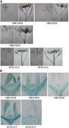
Expression Pattern of UBC1, UBC2, UBC3, HUB1, and HUB2 by Promoter-GUS Fusion. (A) Histochemical analysis of UBC1, UBC2, UBC3, HUB1, and HUB2 expression in leaf, root, and flower. (B) Expression pattern of HUB1, HUB2, and UBCs in the SAMs of 8-d-old seedlings. Nine out of 10 (UBC1), 10 out of 10 (UBC2), 10 out of 11 (UBC3), eight out of 10 (HUB1), and nine out of 10 (HUB2) independent lines exhibited the expression pattern shown. Bar = 10 μm.
Figure 5.
Figure 5.
Physical and Genetic Interactions between HUB1 or HUB2 and UBC1 or UBC2. (A) HUB1 or HUB2 interacts with UBC1 or UBC2. (B) Deletion of the RING finger domain in HUB1 and HUB2 (constructs pGBKT7- HUB1N1-824 and pGADT7-HUB2N1-848) abolishes the interaction between HUB1 or HUB2 and UBC1 or UBC2. Positive control: pGADT7-T + pGBKT7-53; negative control: pGADT7-T + pGBKT7-Lam. The indicated combinations of plasmids were cotransformed into the yeast reporter strain, and the interaction between HUB1,2 and UBCs was assessed by growth on plates lacking Leu, Trp, His, and adenine or by analysis of the activation of β-galactosidase. The full-length and RING finger domain-deleted HUB proteins expressed in the yeast are shown in the bottom. (C) The early-flowering phenotype of hub1 or hub2 and ubc1,2 double and triple mutants. Bar = 1 cm. (D) The rosette leaf number at flowering of the hub1 or hub2 and ubc1,2 double and triple mutants. The data for wild type and each mutant are from at least 20 plants. Error bars indicate sd.
Figure 6.
Figure 6.
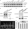
Analysis of the H2Bub1 Level in 7-d-Old Seedlings. (A) Detection of H2Bub1 in wild-type and mutant plants. The upper band was monoubiquitinted Flag-H2B, and the lower band was Flag-H2B. The bands were detected by anti-Flag antibody. H2Bub1, monoubiquitinated H2B; MW, molecular weight. (B) No H2Bub1 was detected in wild-type plants transformed with a mutant form of H2B (K146A). The U in the circle is ubiquitin, and peptide in the bottom of the panel is the C-terminal sequence of H2B. (C) The H2B-specific upper band was recognized by antiubiquitin antibodies after the immunoprecipitation by FLAG antibodies. (D) and (E) Overexpression of the K146A mutant form of histone H2B in wild-type plants (ProH2B:H2BK146A/WT) leads to early flowering. The levels of the mutated form of H2B protein in different lines are shown at the top of the graph in (E). Ten out of 34 independent lines exhibited an early-flowering phenotype (four are shown). Bar in (D) = 1 cm.
Figure 7.
Figure 7.
H2Bub1 Is Required for the Transcriptional Activation of FLC/MAFs. (A) Analysis of the relative expression level of FLC/MAFs by quantitative real-time PCR using RNA from the SAM of 7-d-old seedlings. The levels were normalized to the wild type. (B) Analysis of the relative expression level of FT by quantitative real-time PCR using 10-d-old seedlings. The levels were normalized to the wild type. Three biological replications were performed for each experiment. Actin was used as an internal control. Error bars indicate sd.
Figure 8.
Figure 8.
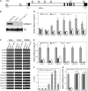
Monoubiquitination of FLC/MAF Chromatin. (A) The structure of FLC. P1 to P8 represent the primers used to assess the level of H2Bub1, H3K4me3, or H3K36me2 by ChIP. The filled boxes represent exons, while the open boxes represent introns. +1, the translation initiation point. (B) Detection of H2Bub1 in Arabidopsis using a monoclonal antibody against H2Bub1. H3 was used as a loading control. (C) ChIP analysis of the H2Bub1 level in FLC/MAFs chromatin followed by PCR. Antibodies used for IP are indicated at top, and plant genotypes are indicated above each lane. (D) ChIP was used to analyze the level of H2Bub1 in chromatin across the FLC locus (P1 to P8 as in [A]), including the promoter and gene body region, from wild-type, hub1, and ubc1 ubc2 seedlings (light gray, dark gray, and white bars, respectively). The amount of immunoprecipitated chromatin as normalized to the total amount of chromatin used in FLCP1 region in wild-type plants was given as 1. (E) Differences in the level of H2Bub1 in the gene body region of FLC/MAF chromatin between wild-type and mutant plants as detected by ChIP analysis. (F) ChIP analysis of the H2Bub1 level in FLC/MAF chromatin from wild-type seedlings. The data represent the amount of immunoprecipitated chromatin versus the total amount of chromatin used. (G) Detection of H2Bub1, H3K4me3, and H3K36me2 in ACTIN chromatin by ChIP analysis. At least three independent immunoprecipitations were performed for each experiment. Unless otherwise indicated, the immunoprecipitated DNA was quantified by quantitative real-time PCR. The data in (E) and (G) were normalized to the input chromatin.
Figure 9.
Figure 9.
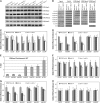
H2Bub1 Is Required for H3K4 and H3K36 Hypermethylation in FLC/MAF Chromatin. (A)Analysis of H3K4, H3K9, and H3K36 methylation on a genome-wide scale by protein gel blotting. (B) Detection of H3K4me3, H3K36me2, and H3K9me2 in the gene body region of FLC/MAF chromatin from wild-type and mutant plants by ChIP analysis followed by PCR. ACTIN was used as an internal control for H3K4me3 and H3K36me2, while TA3 was used as an internal control for H3K9me2. (C) Detection of H3K4me3 in the promoter and gene body regions of FLC chromatin by ChIP analysis. (D) ChIP analysis of the H3K4me3 level in FLC/MAF chromatin. (E) ChIP analysis of the H3K4me3 level in FLC/MAF chromatin from wild-type seedlings. The data represent the amount of immunoprecipitated chromatin versus the total amount of chromatin used. (F) Detection of H3K36me2 in the promoter and gene body regions of FLC chromatin by ChIP analysis. (G)ChIP analysis of the H3K36me2 level in FLC/MAF chromatin. (H)ChIP analysis of the H3K9me2 level in FLC/MAF chromatin. At least three independent immunoprecipitations were performed for each experiment. Unless otherwise indicated, the immunoprecipitated DNA was quantified by quantitative real-time PCR. The data in (C), (D), (F), and (G) were normalized to ACTIN, while the data in (H) were normalized to TA3.
Figure 10.
Figure 10.
Genetic Interaction between HUB1, HUB2, and ELF7. The plants were grown under long-day conditions for 26 d. The rosette leaf number at flowering of the hub1 or hub2 and elf7 double and triple mutants is shown at the bottom. The data for the wild type and each mutant are at least from 20 plants. Error bars indicate sd. Bar = 1 cm.
Figure 11.
Figure 11.
Proposed Model for the Modulation of FLC/MAF Chromatin and Transcription by the UBC1,2/HUB1,2 Pathway in the Control of Flowering Time in Arabidopsis. In this model, HUB1 and HUB2 form a heterotetramer with two HUB1 and two HUB2 molecules to recruit UBC1 or UBC2 to FLC/MAF chromatin, followed by the transfer of ubiquitin from UBC1 or UBC2 to H2B. The ubiquitinated H2B may then serve as a platform for the recruitment of histone H3 methyltransferase for the enhancement of H3K4 and H3K36 hypermethylation, thus leading to increased FLC/MAF expression and the delay of flowering in Arabidopsis.

References

    1. Alonso, J.M., et al. (2003). Genome-wide insertional mutagenesis of Arabidopsis thaliana. Science 301 653–657. - PubMed
    1. Asai, T., Tena, G., Plotnikova, J., Willmann, M.R., Chiu, W.L., Gomez-Gomez, L., Boller, T., Ausubel, F.M., and Sheen, J. (2002). MAP kinase signalling cascade in Arabidopsis innate immunity. Nature 415 977–983. - PubMed
    1. Bastow, R., Mylne, J.S., Lister, C., Lippman, Z., Martienssen, R.A., and Dean, C. (2004). Vernalization requires epigenetic silencing of FLC by histone methlation. Nature 427 164–167. - PubMed
    1. Baurle, I., and Dean, C. (2006). The timing of developmental transition in plants. Cell 125 655–664. - PubMed
    1. Berna, G., Robles, P., and Micol, J.L. (1999). A mutational analysis of leaf morphogenesis in Arabidopsis thaliana. Genetics 152 729–742. - PMC - PubMed

Publication types

MeSH terms