. 2008 Nov;135(21):3577-86.
doi: 10.1242/dev.022350.

Pbx1 functions in distinct regulatory networks to pattern the great arteries and cardiac outflow tract

Affiliations

Pbx1 functions in distinct regulatory networks to pattern the great arteries and cardiac outflow tract

Ching-Pin Chang et al. Development. 2008 Nov.

Abstract

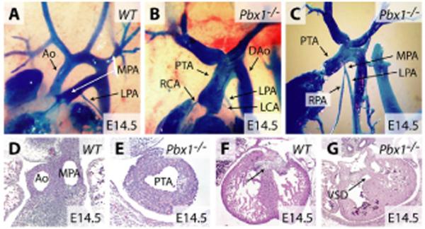
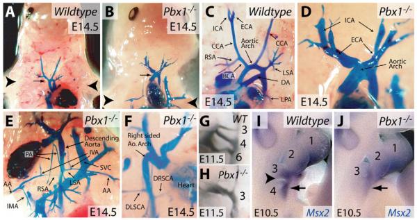
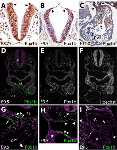
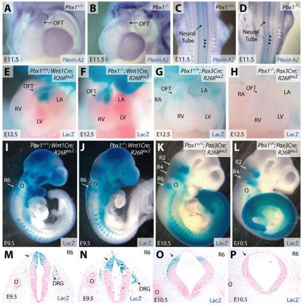
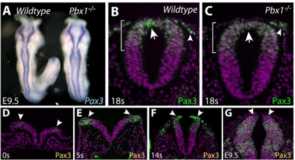
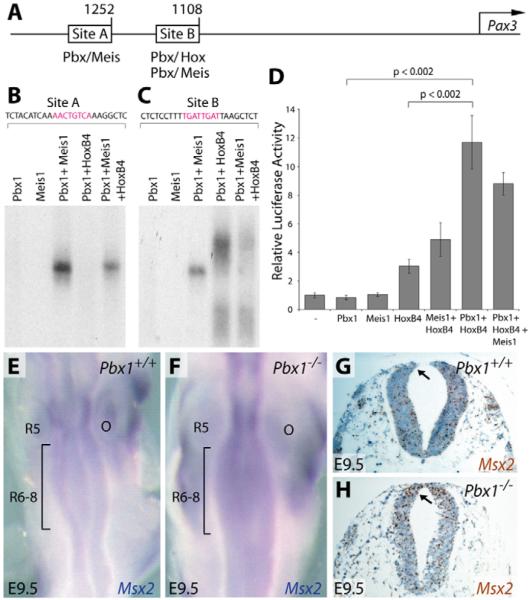
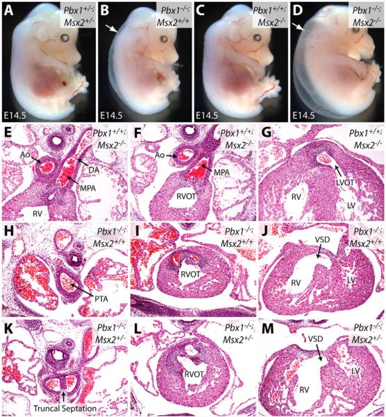
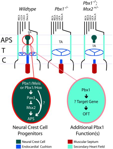
The patterning of the cardiovascular system into systemic and pulmonic circulations is a complex morphogenetic process, the failure of which results in clinically important congenital defects. This process involves extensive vascular remodeling and coordinated division of the cardiac outflow tract (OFT). We demonstrate that the homeodomain transcription factor Pbx1 orchestrates separate transcriptional pathways to control great-artery patterning and cardiac OFT septation in mice. Pbx1-null embryos display anomalous great arteries owing to a failure to establish the initial complement of branchial arch arteries in the caudal pharyngeal region. Pbx1 deficiency also results in the failure of cardiac OFT septation. Pbx1-null embryos lose a transient burst of Pax3 expression in premigratory cardiac neural crest cells (NCCs) that ultimately specifies cardiac NCC function for OFT development, but does not regulate NCC migration to the heart. We show that Pbx1 directly activates Pax3, leading to repression of its target gene Msx2 in NCCs. Compound Msx2/Pbx1-null embryos display significant rescue of cardiac septation, demonstrating that disruption of this Pbx1-Pax3-Msx2 regulatory pathway partially underlies the OFT defects in Pbx1-null mice. Conversely, the great-artery anomalies of compound Msx2/Pbx1-null embryos remain within the same spectrum as those of Pbx1-null embryos. Thus, Pbx1 makes a crucial contribution to distinct regulatory pathways in cardiovascular development.


Figures

Fig. 1
Fig. 1. Pbx1 is required for cardiac OFT septation
(A-C) Angiographic casting of wild-type (A) and Pbx1-/- (B,C) E14.5 mouse embryos. (D-G) Hematoxylin and Eosin-stained transverse sections through the outflow tract (D,E) and ventricular septal regions (F,G) of wild-type (D,F) and Pbx1-/- (E,G) E14.5 embryos. The arrow indicates the interventricular septum. Ao, aorta; MPA, main pulmonary artery; RPA and LPA, right and left pulmonary arteries; PTA, persistent truncus arteriosus; RCA and LCA, right and left coronary arteries; DAo, descending artery; VSD, ventricular septal defect.
Fig. 2
Fig. 2. Pbx1 contributes to patterning of the branchial arch arteries
(A-F) Angiographic casting of wild-type (A,C) and Pbx1-/- (B,D-F) E14.5 mouse embryos. (A,B) The position of the aortic arch (arrow) is indicated relative to the forelimbs (arrowheads). (G,H) India ink casting of the branchial arch arteries in wild-type (G) and Pbx1-/- (H) E11.5 embryos. The branchial arch arteries are numbered according to branchial arch origin. (I,J) Whole-mount in situ hybridization staining for Msx2 (blue) on wild-type (I) and Pbx1-/- (J) E10.5 embryos. The arrowhead indicates the groove separating the third and fourth branchial arches in the wild-type embryo. The arrow indicates Msx2 expression in the fourth branchial arch. CCA, common carotid artery; ICA, internal carotid artery; ECA, external carotid artery; DA, ductus arteriosus; BCA, brachiocephalic artery; RSA and LSA, right and left subclavian arteries; LPA, left pulmonary artery; PA, right and left pulmonary arteries; IVA, internal vertebral artery; AA, axillary artery; IMA, internal mammary artery; SVC, superior vena cava; DLSCA, developmentally left, but anatomically right subclavian artery; DRSCA, developmentally right, but anatomically left subclavian artery.
Fig. 3
Fig. 3. Pbx1 is present in multiple cell types that influence OFT septation and artery patterning, including premigratory neural crest cells
(A-C) Immunohistochemical analysis of the Pbx1b isoform of Pbx1 (brown) in sections of mouse neural tube at E8.75 (A) and E9.5 (B) and the outflow tract (OFT) at E11.0 (C). Sections are counterstained with Hematoxylin. (A,B) Arrowheads indicate the neuroectodermal junction where neural crest cells (NCCs) originate. Arrows indicate paraxial mesodermal cells. (C) Arrowheads show vascular smooth muscle cells of the OFT. The asterisk indicates the endocardial cushion, the arrow cushion endocardium. (D-I) Immunofluorescent staining for Pbx1b in sections of a wild-type E9.5 embryo. (D,G,H,I) Pbx1b is in green and the nuclei are purple (stained with Hoechst). (E,F) The Pbx1b and Hoechst channels are shown separately in grayscale. (G,H). Higher-magnification views of specific regions of the embryo shown in D. (G) Arrowheads indicate secondary heart field and/or NCCs entering the OFT. The arrow points to ectodermal cells. The double arrow indicates endothelial cells of the OFT. (H) The arrowheads indicate endodermal cells lining the pharyngeal pouch. The arrow indicates a paraxial mesodermal cell. The double arrow indicates mesenchymal cells of the branchial arches. (I) Pbx1b immunofluorescent staining of a section of an E9.5 embryonic heart. Arrows mark endocardial cells and arrowheads indicate pericardial cells. RV, right ventricle; NT, neural tube; AS, aortic sac.
Fig. 4
Fig. 4. Pbx1 deficiency has no effect on the migration of cardiac NCCs but abolishes Pax3 proximal promoter activity in rhombomere 6
(A-D) Whole-mount in situ hybridization for plexin A2 transcripts (blue) in Pbx1+/+ (A,C) and Pbx1-/- (B,D) E11.5 mouse embryos. Frontal heart views (A,B) and dorsal views (C,D) of the embryos are shown. Black arrows indicate dorsal root ganglia and white arrows indicate sympathetic chains. (E-L) Whole-mount b-galactosidase (lacZ) staining (blue) of Pbx1+/+ (E,G,I,K) and Pbx1-null (F,H,J,L) embryos of the indicated ages and genotypes. lacZ expression is driven by the Rosa26RlacZ (R26RlacZ) allele in cell lineages that express Cre recombinase driven by either the Wnt1 promoter (Wnt1Cre, E,F,I,J) or the Pax3 1.6 kb proximal promoter (Pax3Cre, G,H,K,L). (M-P) Transverse sections of the embryos shown in I-L at the level of R6. lacZ-expressing cells, derived from progenitors expressing either Wnt1Cre (M,N) or Pax3Cre (O,P), stain blue. Sections are counterstained with Nuclear Fast Red (pink). DRG, dorsal root ganglia; OFT, outflow tract; R, rhombomere; O, otic vesicle; RV, right ventricle; LV, left ventricle; LA, left atrium; RA, right atrium.
Fig. 5
Fig. 5. Pbx1 is required for transient high expression of Pax3 in premigratory NCCs
(A) Dorsal view of wild-type (left) and Pbx1-/- (right) E9.5 mouse embryos stained by whole-mount in situ hybridization for Pax3 transcripts (blue). (B-G) Transverse sections of embryos of the indicated ages [by somite (s) number (between E7.5 and E9.0) or embryonic date] immunostained for Pax3 (green) and with Hoechst (purple). (B,C) Sections through R6 of wild-type (B) and Pbx1-/- (C) 18s embryos. The extent of the Pax3+ domain in the dorsal neural tube is marked by a bracket. Arrows indicate a cluster of premigratory NCCs at the extreme dorsal end of the neural tube. Arrowheads mark migrating NCCs. (D-G) A time course of Pax3 expression in the hindbrain between E7.5 and E9.5. Arrowheads mark the dorsal end of the neural tube.
Fig. 6
Fig. 6. Pbx1 transcriptional complexes activate the Pax3 promoter and are required for repression of Msx2 expression
(A) Schematic of the mouse Pax3 promoter showing the location of Pbx, Meis and Hox binding sites within Sites A and B. Numbers denote the distance (in bp) from the transcription start site (arrow) of the Pax3 gene. (B,C) Electrophoretic mobility shift assays using the indicated in vitro translated transcription factors (Pbx1, Meis1 and HoxB4) and radiolabeled DNA fragments from Site A (B) or Site B (C) of the Pax3 promoter. (D) Transient reporter assays using a transfected plasmid containing the Pax3 1.6 kb proximal promoter driving luciferase expression and co-transfected plasmids expressing the indicated transcription factors. Fold activation was calculated relative to reporter baseline activity following normalization and is presented as the mean ± one s.d. P-values were determined using Student's t-test. (E,F) Whole-mount in situ hybridization for Msx2 (blue) in the hindbrain (dorsal view) in Pbx1+/+ (E) and Pbx1-/- (F) E9.5 embryos. The bracketed regions indicate R6-8. (G,H) Msx2 in situ hybridization (brown) on sections through R6 of Pbx1+/+ (G) and Pbx1-/- (H) E9.5 embryos. The sections are counterstained with Hematoxylin (blue). The arrows indicate premigratory neural crest. O, otic vesicle; R, rhombomere.
Fig. 7
Fig. 7. Msx2 deficiency rescues the truncal septation defect of Pbx1-/- embryos
(A-D) Gross morphology of E14.5 mouse embryos of the indicated genotypes with Pbx1 and Msx2 loss-of-function alleles. Arrows indicate generalized edema. (E-M) Hematoxylin and Eosin-stained transverse sections through E14.5 embryos of the indicated genotypes showing that loss of one allele of Msx2 rescues truncal but not conal septation in Pbx1-/- embryos. Ao, aorta; DA, ductus arteriosus; MPA, main pulmonary artery; RV, right ventricle; RVOT and LVOT, right and left ventricular OFTs; LV, left ventricle; PTA, persistent truncus arteriosus; VSD, ventricular septal defect.
Fig. 8
Fig. 8. A working model of Pbx1 function during branchial arch artery and conotruncal development in the mouse
In the absence of Pbx1, the right and left internal and external carotid arteries branch directly off the aortic arch, the brachiocephalic artery is missing, and the right and left subclavian arteries have aberrant origins from the descending aorta. These defects originate in a failure to establish the fourth and sixth aortic arches owing to small or absent fourth and sixth branchial arches. In premigratory NCCs, Pbx1-Meis and/or Pbx1-Hox transcriptional complexes activate a transient but robust Pax3 expression that is seen on E8. This induction is required to repress Msx2 and ultimately guide establishment of the aorticopulmonary septum by cardiac NCCs. Pbx1 complexes have additional, uncharacterized functions within NCCs or other cells in which Pbx1 is expressed, including those of the secondary heart field, to regulate conotruncal septation at the base of the great arteries (aorta and main pulmonary artery) and conal region of the heart. APS, aorticopulmonary septum; DA, ductus arteriosus; T, truncus; C, conus; TA, truncus arteriosus; RBCA, right brachiocephalic artery; LCCA, left common carotid artery; LSCA, left subclavian artery; Desc.A. descending aorta; LICA and RICA, left and right internal carotid arteries; LECA and RECA, left and right external carotid arteries.

References

    1. Abu-Issa R, Smyth G, Smoak I, Yamamura K, Meyers EN. Fgf8 is required for pharyngeal arch and cardiovascular development in the mouse. Development. 2002;129:4613–4625. - PubMed
    1. Abu-Shaar M, Ryoo HD, Mann RS. Control of the nuclear localization of Extradenticle by competing nuclear import and export signals. Genes Dev. 1999;13:935–945. - PMC - PubMed
    1. Arnold JS, Werling U, Braunstein EM, Liao J, Nowotschin S, Edelmann W, Hebert JM, Morrow BE. Inactivation of Tbx1 in the pharyngeal endoderm results in 22q11DS malformations. Development. 2006;133:977–987. - PubMed
    1. Brendolan A, Ferretti E, Salsi V, Moses K, Quaggin S, Blasi F, Cleary ML, Selleri L. A Pbx1-dependent genetic and transcriptional network regulates spleen ontogeny. Development. 2005;132:3113–3126. - PubMed
    1. Brown CB, Feiner L, Lu MM, Li J, Ma X, Webber AL, Jia L, Raper JA, Epstein JA. PlexinA2 and semaphorin signaling during cardiac neural crest development. Development. 2001;128:3071–3080. - PubMed

Publication types

MeSH terms