. 2008 Dec 12;283(50):34588-95.
doi: 10.1074/jbc.M805928200. Epub 2008 Oct 10.

Active cystathionine beta-synthase can be expressed in heme-free systems in the presence of metal-substituted porphyrins or a chemical chaperone

Affiliations

Active cystathionine beta-synthase can be expressed in heme-free systems in the presence of metal-substituted porphyrins or a chemical chaperone

Tomas Majtan et al. J Biol Chem. .

Abstract

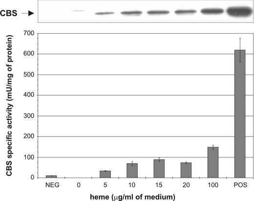
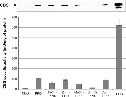
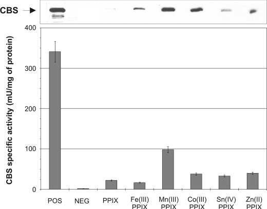
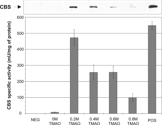
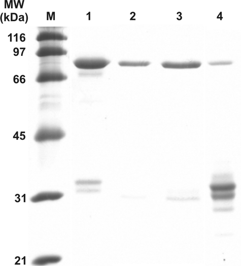
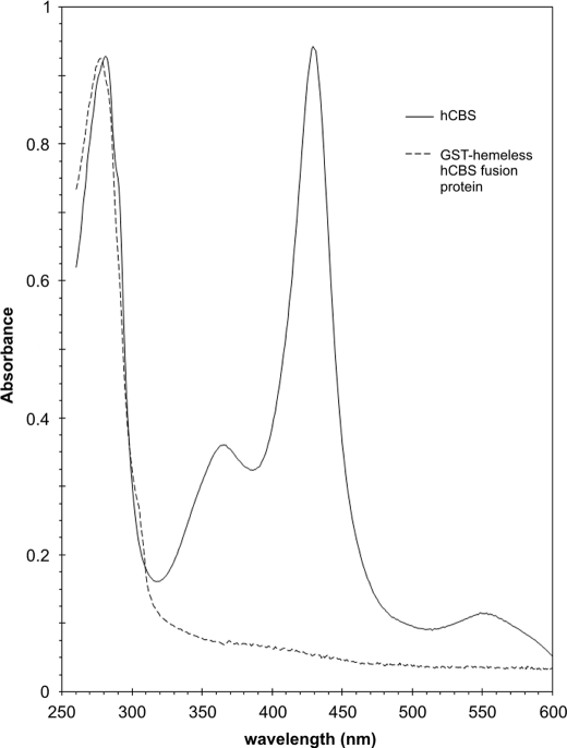
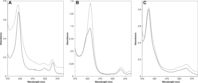
Cystathionine beta-synthase (CBS), a key enzyme in the metabolism of homocysteine, has previously been shown to require a heme co-factor for maximal activity. However, the biochemical function of the CBS heme is not well defined. Here, we show that expression of human CBS in heme-deficient strains of Saccharomyces cerevisiae and Escherichia coli results in production of an enzyme that is misfolded and degraded. Addition of exogenous heme, porphyrins with non-iron metal, or porphyrin lacking metal entirely produced stable and active CBS enzyme. Purification of recombinant CBS enzyme expressed in the presence of various metalloporphyrins confirmed that Mn(III) and Co(III) had 30-60% of the specific activity of Fe(III)-CBS, and still responded to allosteric activation by S-adenosyl-L-methionine. Treatment of S. cerevisiae with the chemical chaperone trimethylamine-N-oxide resulted in near complete restoration of function to human CBS produced in a heme-deficient strain. Taken together, these results suggest that porphyrin moiety of the heme plays a critical role in proper CBS folding and assembly, but that the metal ion is not essential for this function or for allosteric regulation by S-adenosyl-L-methionine.


Figures

FIGURE 1.
FIGURE 1.
Dependence of CBS expression and activity on heme concentration. Western blot of yeast strain WY254 bearing plasmid expressing human CBS. Cell lysates (20 μg/lane) were electrophoresed on a 9% SDS-gel. Following Western transfer, CBS was detected using anti-CBS monoclonal antibody (upper panel). The cell lysates were assayed for CBS activity and the results were plotted (bottom panel). The strains WY61 and WY79 served as positive and negative controls, respectively.
FIGURE 2.
FIGURE 2.
Expression and activity of human CBS in the presence of various heme analogs in yeast system. Cell lysates (20 μg/lane) were electrophoresed on a 9% SDS gel. Following Western transfer, CBS was detected using anti-CBS monoclonal antibody (upper panel). Graph of corresponding CBS activities (bottom panel). Strains WY61 and WY79 served as positive and negative controls, respectively.
FIGURE 3.
FIGURE 3.
Expression and activity of human CBS in the presence of various heme analogs in E. coli system. Western blot of E. coli RP523 bearing plasmid expressing human CBS. Cell lysates (40 μg/lane) were electrophoresed on a 9% SDS gel. Following Western transfer, CBS was detected using anti-CBS monoclonal antibody (upper panel). Graph of corresponding CBS activities (bottom panel).
FIGURE 4.
FIGURE 4.
Impact of chemical chaperone TMAO on human CBS expression and activity in the absence of heme. Western blot of yeast bearing plasmid expressing human CBS. Crude extracts (35 μg/lane) were electrophoresed on a 9% SDS gel. Following Western transfer, CBS was detected using anti-CBS monoclonal antibody (upper panel). Graph of corresponding CBS activities (bottom panel). Strains WY61 and WY79 served as positive and negative controls, respectively.
FIGURE 5.
FIGURE 5.
SDS-PAGE analysis of purified recombinant GST human CBS fusion proteins. 10 μg of each protein was separated on 9% SDS-PAGE gel and stained with SimplyBlue SafeStain (Invitrogen). Lanes: M, broad range SDS-PAGE marker (Bio-Rad); 1, Fe(III)-PPIX hCBS; 2, Mn(III)-PPIX hCBS; 3, Co(III)-PPIX hCBS; 4, GST-hemeless hCBS fusion proteins. The small bands visible in lane 4 when examined on a Western blot are not detectable with anti-CBS or anti-GST antibody.
FIGURE 6.
FIGURE 6.
Absorption spectra of WT human CBS and GST-hemeless hCBS fusion protein. The proteins were diluted to final concentration 0.8 μg/ml in 20 mm HEPES, pH 7.4, 1 mm Tris-(2-carboxyethyl)phosphine, 0.01% Tween 20. Spectra were recorded on HP8453 UV-visible spectrophotometer.
FIGURE 7.
FIGURE 7.
Reduced pyridine hemochromogen spectra of metalloporphyrins extracted from various heme-substituted CBS enzymes. Protoporphyrins were released from the respective CBS proteins (200 μg) in 30 mm NaOH and subsequently stabilized by adding 1 volume of pyridine to 3 volumes of the alkali mixture. Samples were reduced with a trace of solid Na2S2O4 and immediately measured. Solid line represents protoporphyrin released from GST-CBS fusion proteins. Dashed line corresponds to relevant protoporhyrin substance used as a standard. Stock solutions of metalloporhyrins were prepared at 2 mg/ml in 0.2 m sodium phosphate, pH 12. The standards were subsequently prepared the same way as the CBS proteins. A, iron; B, manganese; C, cobalt.

References

    1. Mudd, S. H., Levy, H. L., and Kraus, J. P. (2001) in The Metabolic and Molecular Bases of Inherited Disease (Scriver, C. R., Beaudet, A. L., Sly, W. S., Valle, D., Childs, B., Kinzler, K., and Vogelstein, B., eds) pp. 2007–2056, 8 Ed., McGraw-Hill, New York
    1. Stampfer, M. J., Malinow, M. R., Willett, W. C., Newcomer, L. M., Upson, B., Ullmann, D., Tishler, P. V., and Hennekens, C. H. (1992) J. Am. Med. Assoc. 268 877–881 - PubMed
    1. Kruman, I. I., Kumaravel, T. S., Lohani, A., Pedersen, W. A., Cutler, R. G., Kruman, Y., Haughey, N., Lee, J., Evans, M., and Mattson, M. P. (2002) J. Neurosci. 22 1752–1762 - PMC - PubMed
    1. Mills, J. L., Scott, J. M., Kirke, P. N., McPartlin, J. M., Conley, M. R., Weir, D. G., Molloy, A. M., and Lee, Y. J. (1996) J. Nutr. 126 756S–760S - PubMed
    1. Seshadri, S., Beiser, A., Selhub, J., Jacques, P. F., Rosenberg, I. H., D'Agostino, R. B., Wilson, P. W., and Wolf, P. A. (2002) N. Engl. J. Med. 346 476–483 - PubMed

Publication types

LinkOut - more resources