. 2008 Dec 11;456(7223):819-23.
doi: 10.1038/nature07392. Epub 2008 Oct 12.

DNA double-strand breaks activate a multi-functional genetic program in developing lymphocytes

Affiliations

DNA double-strand breaks activate a multi-functional genetic program in developing lymphocytes

Andrea L Bredemeyer et al. Nature. .

Abstract

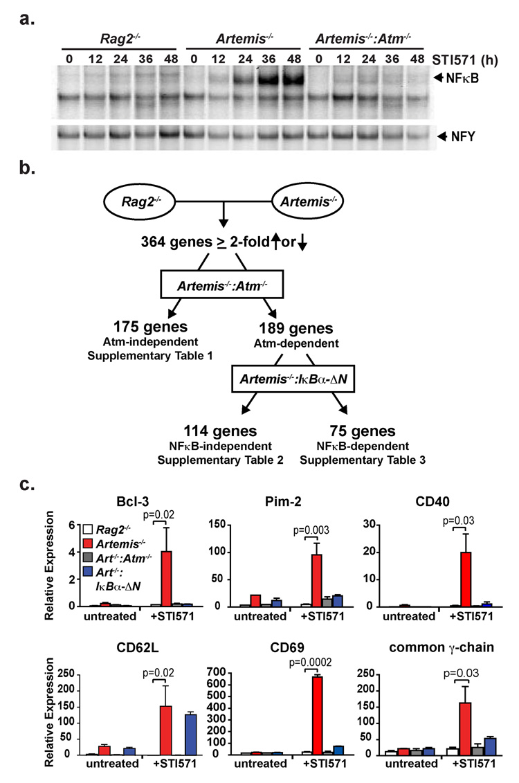
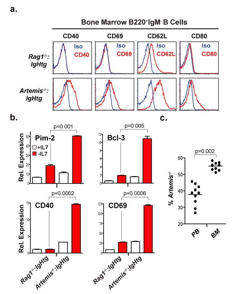
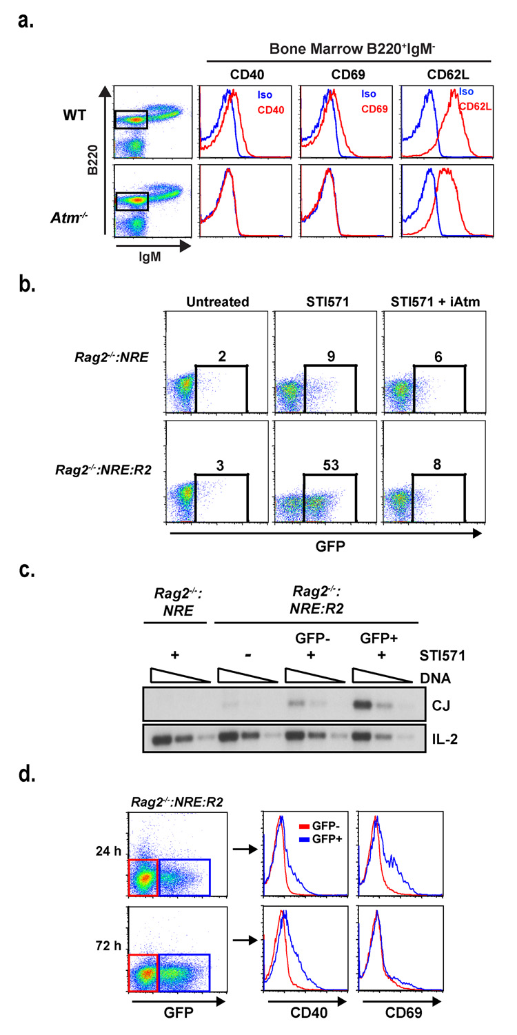
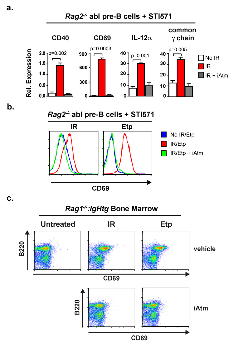
DNA double-strand breaks are generated by genotoxic agents and by cellular endonucleases as intermediates of several important physiological processes. The cellular response to genotoxic DNA breaks includes the activation of transcriptional programs known primarily to regulate cell-cycle checkpoints and cell survival. DNA double-strand breaks are generated in all developing lymphocytes during the assembly of antigen receptor genes, a process that is essential for normal lymphocyte development. Here we show that in murine lymphocytes these physiological DNA breaks activate a broad transcriptional program. This program transcends the canonical DNA double-strand break response and includes many genes that regulate diverse cellular processes important for lymphocyte development. Moreover, the expression of several of these genes is regulated similarly in response to genotoxic DNA damage. Thus, physiological DNA double-strand breaks provide cues that can regulate cell-type-specific processes not directly involved in maintaining the integrity of the genome, and genotoxic DNA breaks could disrupt normal cellular functions by corrupting these processes.


Figures

Figure 1
Figure 1. Rag DSBs activate a broad genetic program
(a) NFκB EMSA of nuclear lysates from Rag2−/−, Artemis−/− and Artemis−/−:Atm−/− abl pre-B cells treated with STI571 for the indicated number of hours. NFY EMSA is shown as a control. Results are representative of three experiments. (b) Schematic of gene expression changes in response to Rag DSBs by microarray analyses. The data are reported in Supplementary Tables 1–3. (c) RT-PCR analysis of mRNA isolated from Rag2−/− (white bars), Artemis−/− (red bars), Artemis−/−:IkBα-ΔN (blue bars) and Artemis−/−:Atm−/− (grey bars) abl pre-B cells treated with STI571 for 0 or 48 hours. Mean and standard deviation from two experiments. P values calculated using a one-tailed t-test.
Figure 2
Figure 2. Rag DSB-dependent gene expression changes in developing B cells in vivo
(a) Flow cytometric analyses of CD40, CD69 CD80 and CD62L expression by B220+:IgM bone marrow B cells (primarily pre-B cells) from Rag1−/−:IgHtg and Artemis−/− :IgHtg mice (gated in Supplementary Fig. 8). Histograms for specific antibodies (red) and isotype controls (blue) are shown. (b) RT-PCR of gene expression in bone marrow cultures derived from Rag1−/−:IgHtg and Artemis−/−:IgHtg mice before (+IL7, white bar) and 48 hours after (−IL7, red bar) the removal of IL7. Mean and standard deviation from two experiments. P values calculated using a one-tailed t-test. (c) Percentage of Artemis−/− abl pre-B cells in the peripheral blood (PB) and bone marrow (BM) of Rag-deficient mice after co-injection of STI571-treated Artemis−/− and Rag-2−/− abl pre-B cells at a 1:1 ratio. The mean (line) of each set of mice analyzed is indicated and P values were calculated using a two-tailed Wilcoxon matched pairs test.
Figure 3
Figure 3. NFκB activation in response to transient Rag DSBs
(a) Flow cytometric analyses of CD40, CD69 and CD62L expression by B220+:IgM bone marrow B cells (gated cells in dot plot) from WT and Atm−/− mice. Histograms for the specific antibodies (red) and isotype control (blue) are shown. (b) Flow cytometric analysis of GFP expression in Rag2−/−:NRE and Rag2−/−:NRE:R2 cells treated with STI571 for 48 hours in the presence or absence of the Atm inhibitor KU-55933 (iAtm). The percentage of GFP+ cells is indicated. Results are representative of three experiments. (c) Rag2−/− :NRE and Rag2−/−:NRE:R2 abl pre-B cells were un-treated (−) or treated with STI571 for 48 hours (+) and GFP+ and GFP Rag2−/−:NRE:R2 abl pre-B cells isolated by flow cytometric cell sorting. Serial 4-fold dilutions of genomic DNA from all cells was assayed for pMX-DELCJ rearrangement by PCR (see Supplementary Fig. 15b). Results are representative of two experiments. (f) Flow cytometric analysis of CD40 and CD69 protein expression on GFP+ (blue histograms) and GFP (red histograms) Rag-2−/− :NRE:R2 cells treated with STI571 for 24 or 72 hours. Results are representative of two experiments.
Figure 4
Figure 4. Genotoxic DSBs promote changes in expression of lymphocyte-specific genes
(a) RT-PCR analysis of gene expression in Rag2−/− abl pre-B cells 2 hours after receiving 0 Gy (white bars) or 4 Gy of IR in the presence (grey bars) or absence (red bars) of the Atm inhibitor KU-55933 (iAtm). All cells were treated with STI571 for 24 hours prior to IR. Results are the mean and standard deviation from two experiments. P values were calculated using a one-tailed t-test. (b) Flow cytometric analysis of CD69 expression on STI571-treated Rag2−/− abl pre-B cells treated with IR as described in (a) or 2 hours after being treated with 5µM etoposide (Etp). Results are representative of three experiments. (c) Flow cytometric analysis of B220 and CD69 expression on bone marrow cells from Rag1−/−:IgHtg mice 2 hours after irradiation (0.5 Gy) or etoposide treatment (5µM) in the presence or absence of the Atm inhibitor KU-55933. Results are representative of two experiments.

References

    1. Shiloh Y. ATM and related protein kinases: safeguarding genome integrity. Nat Rev Cancer. 2003;3(3):155–168. - PubMed
    1. Rouse J, Jackson SP. Interfaces between the detection, signaling, and repair of DNA damage. Science. 2002;297(5581):547–551. - PubMed
    1. Zhou BB, Elledge SJ. The DNA damage response: putting checkpoints in perspective. Nature. 2000;408(6811):433–439. - PubMed
    1. Innes CL, et al. ATM requirement in gene expression responses to ionizing radiation in human lymphoblasts and fibroblasts. Mol Cancer Res. 2006;4(3):197–207. - PubMed
    1. Rashi-Elkeles S, et al. Parallel induction of ATM-dependent pro- and antiapoptotic signals in response to ionizing radiation in murine lymphoid tissue. Oncogene. 2006;25(10):1584–1592. - PubMed

MeSH terms