. 2008 Nov;9(11):1297-306.
doi: 10.1038/ni.1663. Epub 2008 Oct 12.

Interactions among the transcription factors Runx1, RORgammat and Foxp3 regulate the differentiation of interleukin 17-producing T cells

Affiliations

Interactions among the transcription factors Runx1, RORgammat and Foxp3 regulate the differentiation of interleukin 17-producing T cells

Fuping Zhang et al. Nat Immunol. 2008 Nov.

Erratum in

  • Nat Immunol. 2009 Feb;10(2):223

Abstract

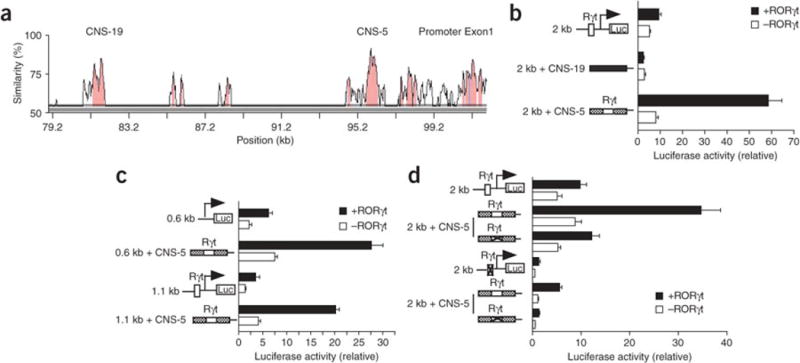
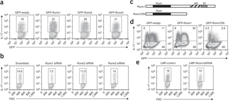
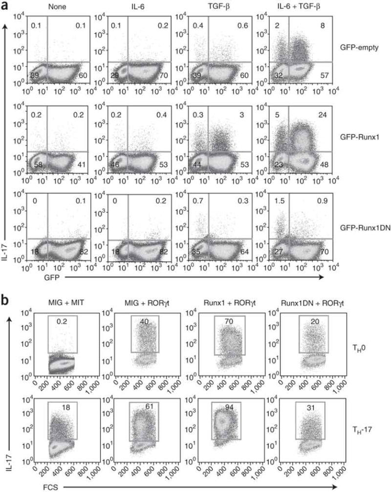
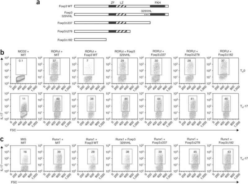
The molecular mechanisms underlying the differentiation of interleukin 17-producing T helper cells (T(H)-17 cells) are still poorly understood. Here we show that optimal transcription of the gene encoding interleukin 17 (Il17) required a 2-kilobase promoter and at least one conserved noncoding (enhancer) sequence, CNS-5. Both cis-regulatory elements contained regions that bound the transcription factors RORgammat and Runx1. Runx1 influenced T(H)-17 differentiation by inducing RORgammat expression and by binding to and acting together with RORgammat during Il17 transcription. However, Runx1 also interacts with the transcription factor Foxp3, and this interaction was necessary for the negative effect of Foxp3 on T(H)-17 differentiation. Thus, our data support a model in which the differential association of Runx1 with Foxp3 and with RORgammat regulates T(H)-17 differentiation.


Figures

Figure 1
Figure 1. Mouse Il17 promoter activity
(a) Luciferase activity of Jurkat cells transfected with fragments (0.6 kb, 1.1 kb and 2 kb) of the mouse Il17 promoter linked to firefly luciferase reporter constructs along with a renillla luciferase vector (transfection efficiency control), allowed to ‘rest’ overnight, then left unstimulated (None) or stimulated for 6 h with PMA and ionomycin (PMA + iono). Results are presented relative to renilla luciferase activity. (b) Luciferase activity of Jurkat cells transfected with luciferase reporter constructs plus RORγt-expressing vector (+RORγt) or empty vector (−RORγt), allowed to ‘rest’ overnight, then stimulated for 6 h with PMA and ionomycin and assessed as described in a. Below, immunoblot analysis of RORγt expression in transfected Jurkat cells, detected with anti-RORγ (α-RORγ). (c) Luciferase activity of Jurkat cells transfected with luciferase reporter constructs containing an intact 2-kb promoter or a 2-kb promoter with a mutated RORγt-binding site, plus RORγt-expressing or empty vector, then stimulated and assessed as described in b. Data are representative of at least four (a,b) or three (c) independent experiments (mean and s.d. of triplicate transfections).
Figure 2
Figure 2. CNS-5 is a RORγt-dependent enhancer element for Il17 transcription
(a) VISTA plot of sequence similarity of CNS sites (pink) in human IL17 and mouse Il17 DNA, presented relative to their positions in the mouse genome (horizontal axis); exons are blue. CNS-19 and CNS-5 (also called CNS2; ref. 14) are located -19 kb and -5 kb, respectively, from the transcriptional start site of mouse Il17. (b) Luciferase activity of Jurkat cells transfected with reporter constructs (left margin) containing the 2-kb promoter alone or linked upstream to CNS-19 (filled rectangles) or CNS-5 (crosshatched rectangles), plus RORγt-expressing or empty vector, stimulated and assessed as described in Figure 1b. Open boxes, RORγt-binding site (Rγt); Luc, luciferase. (c) Luciferase activity of Jurkat cells transfected with luciferase reporter constructs containing 0.6-kb or 1.1-kb promoter fragments alone or linked to CNS-5, plus RORγt-expressing or empty vector, stimulated and assessed as described in Figure 1b. (d) Luciferase activity of Jurkat cells transfected with reporter constructs containing the 2-kb promoter fragment alone or linked to CNS-5, with (‘X’ in box) or without (open box) mutation of RORγt-binding sites, plus RORγt-expressing or empty vector, stimulated and assessed as described in Figure 1b. Data are representative of experiments repeated at least three times (mean and s.d. of triplicate transfections).
Figure 3
Figure 3. Runx1 upregulates Il17 transcription
(a) Luciferase activity of Jurkat cells transfected with a reporter construct containing the 2-kb Il17 promoter fragment, plus Flag-tagged Runx1-expressing or empty vector and a renilla luciferase plasmid, stimulated and assessed as described in Figure 1a. Below, immunoblot analysis of Runx1 expression in transfected Jurkat cells. (b) Luciferase activity of Jurkat cells transfected with reporter constructs containing the 2-kb Il17 promoter fragment with or without mutation of the RORγt-binding site or Runx1-binding sites (Rx), plus RORγt-expressing or empty vector, stimulated and assessed as described in Figure 1b. (c) Luciferase activity of Jurkat cells transfected with reporter constructs containing the 2-kb Il17 promoter fragment with or without mutation of the RORγt-binding site, plus Runx1-expressing vector (+) or empty vector (−), stimulated and assessed as described in Figure 1a. Data are representative of three independent experiments (mean and s.d. of triplicate transfections).
Figure 4
Figure 4. Runx1 is required for IL-17 expression in CD4+ T cells
(a,b) Flow cytometry of IL-17 expression in purified CD4+ cells transduced with the MSCV-IRES-GFP retroviral vector (MIG) encoding GFP alone, GFP-Runx1, GFP-Runx2 or GFP-Runx3 constructs (a) or transfected by nucleofection with Runx1-, Runx2- or Runx3-specific siRNA or control (‘scrambled’) siRNA (b), then cultured in TH-17-polarizing conditions, followed by intracellular staining 4 d after the first transduction (5 d after activation). Plots are gated on GFP+ cells. Numbers in outlined areas indicate percent IL-17+ cells. FSC, forward scatter. Data are representative of three independent experiments. (c) Runx1 and the Runx1DN construct, including the Runt domain, activation domain (AD) and inhibition domain (ID). (d) Flow cytometry of IL-17 production by purified CD4+ T cells transduced with retroviral vectors encoding GFP alone, GFP-Runx1 or GFP-Runx1DN, then cultured in TH-17-polarizing conditions and stained 4 d after the first transduction (5 d after activation). Numbers in quadrants indicate percent cells in each. Data are representative of at least three independent experiments. (e) Flow cytometry of IL-17 in purified naive CD4+ cells transduced with retroviral vector containing control shRNA (LMP control) or Runx1-specific shRNA (LMP Runx1-shRNA), then cultured in TH-17-polarizing conditions and stained on day 5. Plots are gated on GFP+ cells. Numbers in outlined areas indicate percent IL-17+ cells. Data are representative of three independent experiments.
Figure 5
Figure 5. Runx1 acts together with RORγt to induce IL-17 production
(a) Flow cytometry of IL-17 production by purified naive CD4+ T cells transduced with retroviruses as described in Figure 4d and then cultured in the presence of various cytokines (above plots), followed by intracellular staining on day 4 after the first transduction (day 5 after activation). Numbers in quadrants indicate percent cells in each. (b) Flow cytometry of IL-17 expression in purified naive CD4+ T cells transduced with various combinations of retrovirus expressing GFP alone (MIG), Thy-1.1 alone (MIT), Thy-1.1-RORγt, GFP-Runx1 or GFP-Runx1DN, then cultured in TH0- or TH-17-polarizing conditions and analyzed as described in Figure 4a. Plots are gated on GFP+Thy-1.1+ cells. Numbers in outlined areas indicate percent IL-17+ cells. Data are representative of at least three independent experiments.
Figure 6
Figure 6. RORγt and Runx1 bind to the Il17 promoter and enhancer
(a,b) Transcription factor binding in purified CD4+ T cells transduced with retrovirus expressing GFP-Myc-RORγt (a) or GFP-Flag-Runx1 (b) as described in Figure 4a, then cultured for 4 d in TH-17-polarizing conditions and restimulated for 4 h with PMA and ionomycin, and then assessed by ChIP with anti-Myc (a) or anti-Flag (b). (c,d) Binding of endogenous transcription factors in purified CD4+ T cells activated and cultured for 4 d in TH-17-polarizing conditions and restimulated for 4 h with PMA and ionomycin, then assessed by ChIP with anti-RORγ (c) or anti-Runx1 (d). Results are presented as the copy number of genomic DNA detected by real-time PCR in each immunoprecipitation relative to a standard dilution of input DNA. Data are representative of three independent experiments (mean and s.d. of triplicate immunoprecipitations for each antibody).
Figure 7
Figure 7. Runx1 promotes RORγt expression
(a) Real-time RT-PCR of the expression of RORγt and IL-17 in purified CD4+ T cells activated and transduced with retrovirus expressing GFP alone, GFP-Runx1 or GFP-Runx1DN, then cultured in TH-17-polarizing conditions; GFP+ cells were then purified by flow cytometry, recultured overnight in TH-17-polarizing conditions, washed and then restimulated for 6 h with anti-CD3 plus anti-CD28. (b) Real-time RT-PCR of the expression of Runx1, RORγt and IL-17 in purified CD4+ cells transfected with scrambled siRNA or Runx1-specific siRNA as described in Figure 4b, then cultured for 48 h in TH-17-polarizing conditions. Results are presented relative to HPRT expression. Data are representative of three independent experiments (mean and s.d. of triplicates).
Figure 8
Figure 8. Interaction of Runx1, RORγt and Foxp3 regulates TH-17 cell differentiation
(a) Intact Foxp3 (wild-type (WT)) and mutant Foxp3 constructs, including the location of the zinc finger (ZF), leucine zipper (LZ) and forkhead (FKH) domains. Foxp3 329VHL contains the substitutions D329V, Y330H and K332L; Δ337, Δ278 and Δ182 indicate carboxy-terminal deletions (number indicates position at which deletion ends). (b) Flow cytometry of purified naive CD4+ cells activated and transduced as described in Figure 5b with various combinations of the MSCV-IRES-human CD2 (MCD2) retrovirus encoding human CD2 alone, MSCV–IRES–Thy-1.1 (MIT) encoding Thy-1.1 alone, human CD2–RORγt, Thy-1.1–Foxp3 or Thy-1.1–mutant Foxp3, then cultured in TH-17- or TH0-polarizing conditions and analyzed 4 d after transduction. Plots are gated on human CD2–positive Thy-1.1+ cells. (c) Flow cytometry of purified naive CD4+ T cells activated and transduced as described in Figure 5b with various combinations of retrovirus encoding GFP alone (MIG), Thy-1.1 alone (MIT), GFP-Runx1, Thy-1.1–Foxp3 or Thy-1.1–mutant Foxp3, then cultured in TH-17-polarizing conditions and analyzed 4 d after transduction. Plots are gated on GFP+Thy-1.1+ cells. Numbers in outlined areas (b,c) indicate percent IL-17+ cells. Data are representative of at least three independent experiments.
Figure 9
Figure 9. RORγt binds to both Runx1 and Foxp3
(a) Immunoblot (IB) analysis of Runx1 and RORγt in 293T cells transfected with Flag-Runx1 and Myc-RORγt constructs with or without ethidium bromide (EtBr), detected in lysates immunoprecipitated (IP) with anti-Flag or anti-Myc. (b) Immunoassay of purified CD4+ T cells cultured for 4 d in TH-17-polarizing conditions; lysates prepared in the presence of ethidium bromide were immunoprecipitated with IgG isotype-matched control antibody or anti-Runx1, then analyzed by immunoblot with anti-RORγt or anti-Runx1. (c,d) Immunoassay of 293T cells transfected with Myc-tagged RORγt and Flag-tagged wild-type Foxp3 (c,d) or mutant Foxp3 (d); lysates immunoprecipitated with anti-Flag or anti-Myc were analyzed by immunoblot with anti-Flag or anti-Myc. Results are representative of three independent experiments.

Similar articles

Cited by

References

    1. Ivanov II, et al. The orphan nuclear receptor RORγt directs the differentiation program of proinflammatory IL-17+ T helper cells. Cell. 2006;126:1121–1133. - PubMed
    1. Nakae S, Nambu A, Sudo K, Iwakura Y. Suppression of immune induction of collagen-induced arthritis in IL-17-deficient mice. J Immunol. 2003;171:6173–6177. - PubMed
    1. Komiyama Y, et al. IL-17 plays an important role in the development of experimental autoimmune encephalomyelitis. J Immunol. 2006;177:566–573. - PubMed
    1. Wong CK, et al. Proinflammatory cytokines (IL-17, IL-6, IL-18 and IL-12) and Th cytokines (IFN-γ, IL-4, IL-10 and IL-13) in patients with allergic asthma. Clin Exp Immunol. 2001;125:177–183. - PMC - PubMed
    1. Yang XO, et al. Molecular antagonism and plasticity of regulatory and inflammatory T cell programs. Immunity. 2008;29:44–56. - PMC - PubMed

MeSH terms

Substances