Review
. 2008 Oct 13:3:27.
doi: 10.1186/1750-1172-3-27.

Transposition of the great arteries

Affiliations
Review

Transposition of the great arteries

Paula Martins et al. Orphanet J Rare Dis. .

Abstract

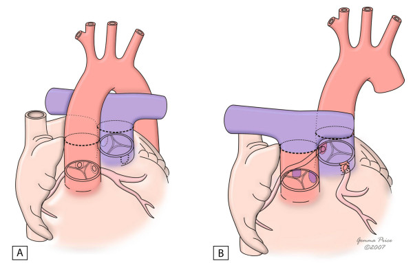
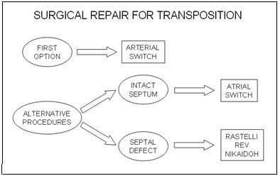
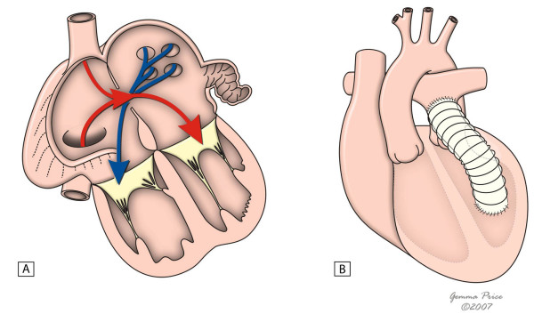
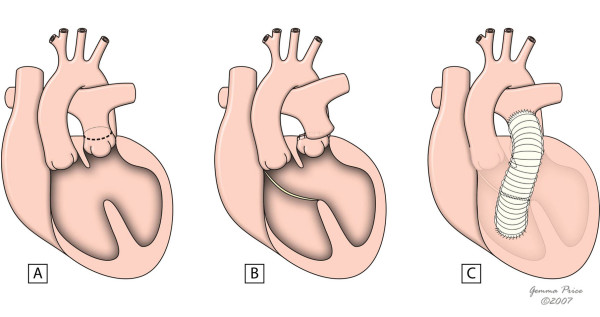
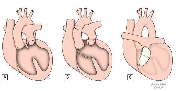
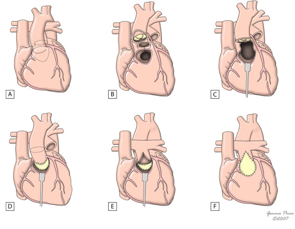
Transposition of the great arteries (TGA), also referred to as complete transposition, is a congenital cardiac malformation characterised by atrioventricular concordance and ventriculoarterial (VA) discordance. The incidence is estimated at 1 in 3,500-5,000 live births, with a male-to-female ratio 1.5 to 3.2:1. In 50% of cases, the VA discordance is an isolated finding. In 10% of cases, TGA is associated with noncardiac malformations. The association with other cardiac malformations such as ventricular septal defect (VSD) and left ventricular outflow tract obstruction is frequent and dictates timing and clinical presentation, which consists of cyanosis with or without congestive heart failure. The onset and severity depend on anatomical and functional variants that influence the degree of mixing between the two circulations. If no obstructive lesions are present and there is a large VSD, cyanosis may go undetected and only be perceived during episodes of crying or agitation. In these cases, signs of congestive heart failure prevail. The exact aetiology remains unknown. Some associated risk factors (gestational diabetes mellitus, maternal exposure to rodenticides and herbicides, maternal use of antiepileptic drugs) have been postulated. Mutations in growth differentiation factor-1 gene, the thyroid hormone receptor-associated protein-2 gene and the gene encoding the cryptic protein have been shown implicated in discordant VA connections, but they explain only a small minority of TGA cases.The diagnosis is confirmed by echocardiography, which also provides the morphological details required for future surgical management. Prenatal diagnosis by foetal echocardiography is possible and desirable, as it may improve the early neonatal management and reduce morbidity and mortality. Differential diagnosis includes other causes of central neonatal cyanosis. Palliative treatment with prostaglandin E1 and balloon atrial septostomy are usually required soon after birth. Surgical correction is performed at a later stage. Usually, the Jatene arterial switch operation is the procedure of choice. Whenever this operation is not feasible, adequate alternative surgical approach should be implemented. With the advent of newer and improved surgical techniques and post operative intensive care, the long-term survival is approximately 90% at 15 years of age. However, the exercise performance, cognitive function and quality of life may be impaired.


Figures

Figure 1
Figure 1
This subcostal view shows the left ventricle originating a vessel that bifurcates, which is thus identified as the pulmonary artery.
Figure 2
Figure 2
Subcostal view showing discordant ventriculoarterial connections together with the presence of parallel, rather than crossing, great arteries arising form the ventricles.
Figure 3
Figure 3
Short axis view showing the aorta giving rise to the coronary arteries (arrow) in an anterior position and to the right. The pulmonary trunk is placed in a central position.
Figure 4
Figure 4
Echocardiography of a complex transposition with a ventricular septal defect and pulmonary stenosis.
Figure 5
Figure 5
The figure illustrates the arterial switch procedure. It shows the connection of the proximal great arteries to the distal end of the other great artery, as well as the transfer of coronary arteries to the new aorta. (adapted from [10]. Martins P, Tran V, Price G, Tsang V and Cook A, Cardiol Young 2008; 18:124–34, with permission from Cambridge University Press).
Figure 6
Figure 6
Surgical procedures used in the correction of the transposition of the great arteries.
Figure 7
Figure 7
This figure shows the common principle subjacent to intra-atrial switch. Figure 7A shows in diagrammatic fashion, the redirection of pulmonary and systemic venous flows at atrial level. An external conduit (B) is placed to overcome the obstruction within the left ventricular outflow tract. (adapted from [10]. Martins P, Tran V, Price G, Tsang V and Cook A, Cardiol Young 2008; 18:124–34, with permission from Cambridge University Press).
Figure 8
Figure 8
The figure shows the main steps of Rastelli procedure in the setting of a deviated outlet septum (A). A patch is placed to create an interventricular tunnel (B), and an extracardiac conduit is placed between the right ventricle and the pulmonary arteries (C). (adapted from [10]. Martins P, Tran V, Price G, Tsang V and Cook A, Cardiol Young 2008; 18:124–34, with permission from Cambridge University Press).
Figure 9
Figure 9
This figure shows the innovations of the REV procedure relative to the Rastelli procedure, with resection of the muscular outlet septum (B) and use of Lecompte manoeuvre (C) which avoids the use of an extacardiac conduit. (adapted from [10]. Martins P, Tran V, Price G, Tsang V and Cook A, Cardiol Young 2008; 18:124–34, with permission from Cambridge University Press).
Figure 10
Figure 10
The figure shows the major issues of Nikaidoh's procedure. Figure 10A shows the areas for harvesting the aortic root and transection of the pulmonary trunk. In 10B, the aorta and pulmonary trunk have already been disconnected from their previous insertion, and the line of division of the outlet septum is shown. Figures 10C and 10D show aortic translocation and reconstruction of the left outflow tract, with a pericardial patch used to close the ventricular septal defect. In 10E and 10F, the pulmonary trunk is sutured to the right ventricular outflow tract, and the reconstruction is completed with a pericardial patch. (adapted from [10]. Martins P, Tran V, Price G, Tsang V and Cook A, Cardiol Young 2008; 18:124–34, with permission from Cambridge University Press).

References

    1. Ho SY, Baker EJ, Rigby ML, Anderson RH. Color Atlas of Congenital Heart Disease – Morphologic and Clinical Correlations. Mosby-Wolfe, London; 1995.
    1. Wilkinson JL, Cochrane AD, Karl TR. Congenital Heart Surgery Nomenclature and Database Project: corrected (discordant) transposition of the great arteries (and related malformations) Ann Thorac Surg. 2000;69:S236–48. doi: 10.1016/S0003-4975(99)01246-1. - DOI - PubMed
    1. Jaggers JJ, Cameron DE, Herlong JR, Ungerleider RM. Congenital Heart Surgery Nomenclature and Database Project: transposition of the great arteries. Ann Thorac Surg. 2000;69:S205–35. doi: 10.1016/S0003-4975(99)01282-5. - DOI - PubMed
    1. Houyel L, Van Praagh R, Lacour-Gayet F, Serraf A, Petit J, Bruniaux J, Planché C. Transposition of the great arteries [S, D, L]. Pathologic anatomy, diagnosis, and surgical management of a newly recognized complex. J Thorac Cardiovasc Surg. 1995;110:613–24. doi: 10.1016/S0022-5223(95)70092-7. - DOI - PubMed
    1. Samánek M, Slavík Z, Zborilová B, Hrobonová V, Vorísková M, Skovránek J. Prevalence, treatment, and outcome of heart disease in live-born children: a prospective analysis of 91,823 live-born children. Pediatr Cardiol. 1989;10:205–11. doi: 10.1007/BF02083294. - DOI - PubMed