. 2008 Oct 10;32(1):70-80.
doi: 10.1016/j.molcel.2008.08.018.

Dif1 is a DNA-damage-regulated facilitator of nuclear import for ribonucleotide reductase

Affiliations

Dif1 is a DNA-damage-regulated facilitator of nuclear import for ribonucleotide reductase

Yang David Lee et al. Mol Cell. .

Abstract

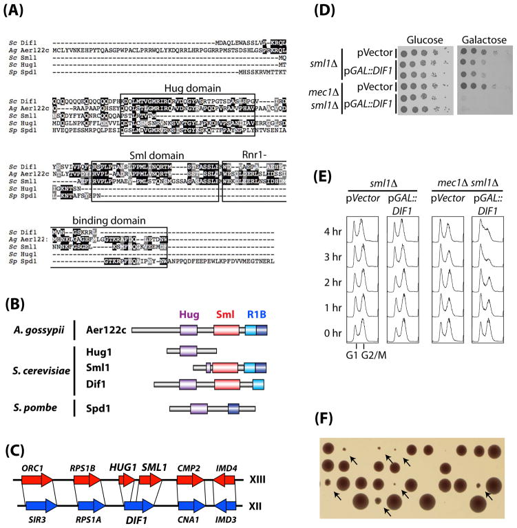
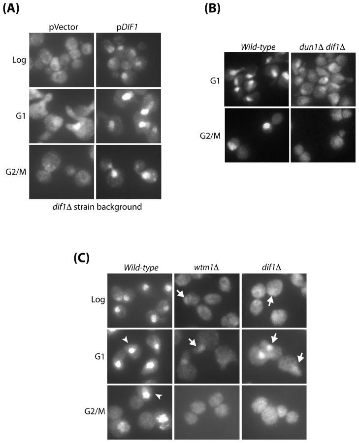
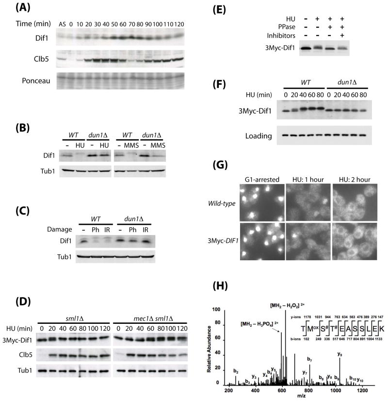
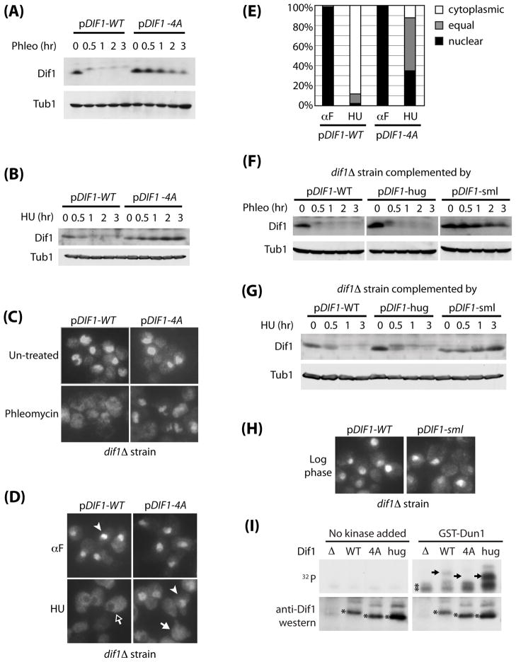
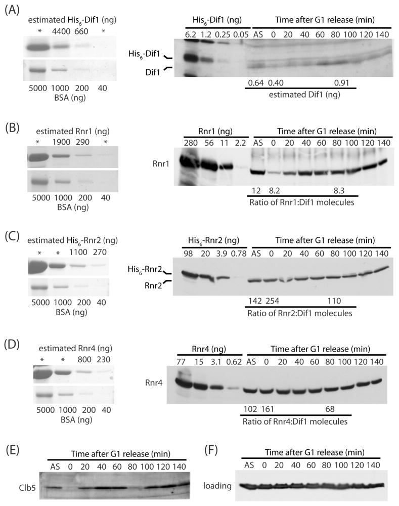
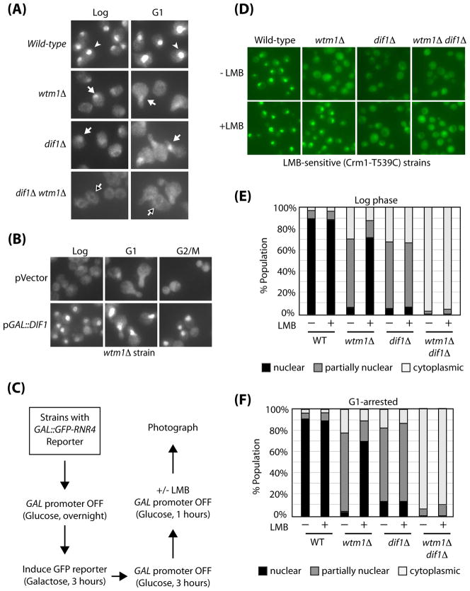
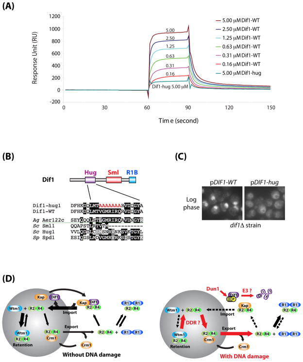
The control of dNTP concentrations is critical to the fidelity of DNA synthesis and repair. One level of regulation is through subcellular localization of ribonucleotide reductase. In Saccharomyces cerevisiae, the small subunit Rnr2-Rnr4 is nuclear, whereas the large subunit Rnr1 is cytoplasmic. In response to S phase or DNA damage, Rnr2-Rnr4 enters the cytoplasm to bind Rnr1, forming an active complex. We previously reported that Wtm1 anchors Rnr2-Rnr4 in the nucleus. Here, we identify DIF1, which regulates localization of Rnr2-Rnr4. Dif1 binds directly to the Rnr2-Rnr4 complex through a conserved Hug domain to drive nuclear import. Dif1 is both cell-cycle and DNA-damage regulated, the latter of which occurs via the Mec1-Dun1 pathway. In response to DNA damage, Dun1 directly phosphorylates Dif1, which both inactivates and degrades Dif1 and allows Rnr2-Rnr4 to become cytoplasmic. We propose that Rnr2-Rnr4 nuclear localization is achieved by a dynamic combination of Wtm1-mediated nuclear retention to limit export and regulated nuclear import through Dif1.


Figures

Figure 1
Figure 1. Dif1 is an inhibitor of S-phase cell cycle progression and a paralog of Sml1
(A) Sequence alignment between S. cerevisiae (Sc) Dif1, Sml1, Hug1, and S. pombe (Sp) Spd1, and A. gossypii (Ag) Aer122c. The three conserved domains: the Hug domain, the Sml domain, and the Rnr1-binding domain, are boxed. (B) Simplified diagrams showing the three different domains of the orthologs of Dif1. The Rnr1-binding domain (R1B) domain, which has been characterized through mutational analysis, was further divided into an N-terminal (cyan) and a C-terminal (blue) sub-domains. (C) Synteny in the proximity of the HUG1/SML1 loci on chromosome XIII and the DIF1 (YLR437c) locus on chromosome XII in S. cerevisiae. (D) sml1Δ and mec1Δ sml1Δ strains containing vector alone or a galactose-inducible DIF1 plasmid (pGAL1::DIF1) were serially diluted and spotted on glucose and galactose media. (E) Cell cycle profiles of sml1Δ and mec1Δ sml1Δ strains over-expressing DIF1. Cells were grown to log phase in glucose before being switched to galactose. Samples were taken from 0 to 4 hours after the galactose switch for FACS analysis of DNA content. (F) Tetrad dissection of MEC1/mec1Δ::his5+ DIF1/dif1Δ::TRP1 diploid. Arrows mark small colonies, which are HU-sensitive and tryptophan- and histidine-prototrophic (data not shown).
Figure 2
Figure 2. Dif1 is required for the nuclear localization of Rnr small subunits
(A) dif1Δ strains complemented with either vector alone or with a plasmid containing the wild-type DIF1 gene were grown to log-phase, or arrested in G1 with α-factor, or arrested in G2/M with nocodazole. Cells were processed for Rnr2 visualization by indirect immunofluorescence. (B) G1- or G2/M-arrested wild-type and dun1Δ dif1Δ strains were processed for Rnr2 visualization by indirect immunofluorescence. (C) Comparison of Rnr2 localization in wild-type, wtm1Δ, and dif1Δ mutants. Cells were either grown to log phase, arrested in G1 or arrested in G2/M. Samples were processed for Rnr2 visualization by indirect immunofluorescence. Examples of cells with strong nuclear Rnr2 staining are marked with white arrowheads, and cells with partially nuclear Rnr2 staining are marked with white arrows.
Figure 3
Figure 3. Dif1 is regulated during cell cycle and DNA damage response
(A) Cell cycle analysis of Dif1 abundance. Cells arrested in G1 by alpha factor were released into the cell cycle. Samples were collected at 10-minute intervals for western blotting of Dif1. Clb5 was used to monitor cell cycle progression. Ponceau staining served as a loading control. A log-phase sample from an asynchronous (AS) population of cells was loaded in the first lane for comparison. (B) Western blot analysis of Dif1 from log-phase wild-type (WT) and dun1Δ cells treated with HU (150 mM, 1.5 hours) or MMS (0.1%, 1.5 hours). Tubulin (Tub1) serves as a loading control. (C) Western blot analysis of Dif1 from wild-type (WT) and dun1Δ cells treated with phleomycin (Ph, 50 ng/mL, 1 hour) or ionizing radiation (IR, 20 kRad, 1-hour irradiation) while maintained in G2/M-arrest. (D) Cell cycle-dependent post-translational modification of 3Myc-Dif1 in sml1Δ and mec1Δ sml1Δ cells. Cells with a single copy of 3Myc-DIF1 integrated at the endogenous locus were arrested in G1 by α-factor, and then released into the cell cycle in the presence of HU (150 mM). Samples were collected at 20-minute intervals for western analysis of 3Myc-Dif1 mobility and abundance. Clb5 was used to monitor cell cycle progression. (E) Phosphatase (PPase) treatment of 3Myc-Dif1. Log-phase yeast cells carrying an integrated 3Myc-DIF1 were treated with HU (150 mM, 1.5 hours). 3Myc-Dif1 was immunoprecipitated from the lysate, and subjected to phosphatase treatment (PPase), with or without phosphatase inhibitors. (F) Dun1-dependent Dif1 phosphorylation after HU-treatment. Wild-type and dun1Δ strains with an integrated 3Myc-DIF1 were arrested in G1 by α-factor, and then released into HU media (150 mM). Samples were collected at 20-minute intervals for western analysis of 3Myc-Dif1 mobility and abundance. (G) Rnr2 visualization by indirect immunofluorescence of wild-type or a 3Myc-DIF1 integrated strain arrested in G1-phase with α-factor, and then released into HU media (150 mM) for 1 and 2 hours. (H) Phospho-mapping of 3Myc-Dif1. Log phase cells with an integrated 3Myc-DIF1 were treated with HU (150 mM, 1.5 hours). 3Myc-Dif1 was immunoprecipitated with anti-Myc antibodies, resolved and silver stained on SDS-PAGE, and excised for phospho-mapping by mass spectrometry. The “#” signs indicate two potential sites for phosphorylation.
Figure 4
Figure 4. The Sml and Hug domains have distinct roles in Dif1 regulation
(A) Western blot analysis of wild-type and phospho-mutant Dif1 after phleomycin treatment. dif1Δ strains containing a wild-type DIF1 plasmid (pDIF1-WT), or the phospho-mutant plasmid (pDIF1-4A) were treated with phleomycin (50 ng/mL) for the indicated times during G2/M arrest by nocodazole. Tubulin (Tub1) serves as a loading control. (B) Western blot analysis of wild-type and phospho-mutant Dif1 from cells arrested in G1 by α-factor, and then released into HU-media (150 mM) for the indicated times. (C) G2/M-arrested wild-type and phospho-mutant Dif1 strains were treated with phleomycin for 2 hours and processed for Rnr2 visualization by indirect immunofluorescence. (D) Wild-type or phospho-mutant Dif1 cells were arrested in G1 by alpha-factor (αF) or released from G1 into HU (150 mM) for 1 hour and processed for Rnr2 visualization by indirect immunofluorescence. Examples of cells with Rnr2 staining that is predominately nuclear (white arrowhead), predominately cytoplasmic (black arrow), or equal in distributions (white arrow) are indicted. (E) Quantification of part D. For each treatment, more than 160 cells were counted (range: 161–200 cells). (F) Western blot analysis of wild-type, hug-domain mutant, or sml-domain mutant Dif1 after phleomycin treatment. dif1Δ strains complemented by plasmids containing wild-type (pDIF1-WT), hug-domain mutated (pDIF1-hug), or sml-domain mutated (pDIF1-sml) versions of DIF1 were treated with phleomycin (50 ng/mL) for the indicated times while maintained in G2/M. Tubulin (Tub1) serves as the loading control. (G) Western blot analysis of Dif1, Dif1-hug, Dif1-sml mutants after HU treatment. dif1Δ strains complemented by plasmids containing wild-type (pDIF1-WT), hug-domain mutant (pDIF1-hug), or sml-domain mutant (pDIF1-sml) versions of DIF1 were arrested in G1 by alpha-factor, and then released into HU media (150 mM) for the indicated times. (H) Rnr2 visualization by indirect immunofluorescence from log phase cultures of dif1Δ strains carrying pDIF1-WT or pDIF1-sml plasmids. (I). Dun1 in vitro kinase assay. Polyclonal anti-Dif1 antibodies were used to immuno-precipitate Dif1 from dif1Δ yeast strain complemented by plasmid alone (Δ), wild-type (WT), phospho-mutant (4A), and hug-domain mutant (hug) Dif1, and then incubated with or without recombinant GST-Dun1 purified from insect cells in the presence of [γ32-P] ATP. Bottom panel shows a western blot with anti-Dif1 antibodies. The predominant Dif1 bands are marked with single asterisks. The difference in their size is due to site-directed mutagenesis. Top panel shows autoradiograph, with the expected position of each Dif1 from the western blot marked by arrows. The non-specific bands present even in the dif1Δ sample was marked by double asterisks.
Figure 5
Figure 5. Dif1 is much less abundant than the Rnr2-Rnr4 heterodimer
(A) Left panel: known standards of bovine serum albumin (BSA) were resolved on SDS-PAGE alongside of five-fold serial dilutions of purified recombinant His6-Dif1 to estimate the concentration of the original Dif1 sample in nanogram. Dif1 serial dilutions that are beyond the range of the BSA standard were not used for estimation of protein concentration and were marked by asterisks (*). Right panel: known quantities of His6-Dif1 were resolved on SDS-PAGE alongside of lysates from asynchronous (AS) and synchronized wild-type cells released from a G1-block and taken at 20-minute intervals. The membrane was probed with anti-Dif1 antibodies. (B) As in part A except measuring Rnr1. Right panel: estimated Rnr1:Dif1 ratios are indicated below the western blot for the asynchronous, 0-minute, and 80-minutes time points. (C) As in part A except measuring Rnr2. (D) As in part A except measuring Rnr4. (E) Clb5 western blot to show progression of S-phase in the synchronized culture. (F) Tubulin western blot serves as loading control.
Figure 6
Figure 6. Dif1 controls the nuclear import of Rnr2-Rnr4
(A) The nuclear localization of Rnr2 in the dif1Δ wtm1Δ double mutant was compared to wild-type, wtm1Δ and dif1Δ single mutants. Log-phase and G1-arrested cells were processed for visualization of Rnr2 by indirect immunofluorescence. Cells with strong nuclear Rnr2 staining (white arrowhead), residual nuclear Rnr2 staining (white arrow), and Rnr2 nuclear exclusion staining (black arrow) are indicated. (B) wtm1Δ strains containing vector alone or a galactose-inducible DIF1 plasmid (pGAL::DIF1) were grown to log phase in glucose media. Cells were then switched to galactose media for 3 hours, while either being kept in log phase or arrested in G1 or G2/M. Samples were processed for Rnr2 visualization by indirect immunofluorescence. (C) A schematic of the experiment used to examine nuclear import of Rnr2-Rnr4. Cells were grown to log-phase in raffinose-glucose media, then switched to raffinose-galactose media for 3 hours to turn on the transcription of the GAL1-GFP-RNR4 reporter. The promoter was shut off by switching back to glucose media for 3 hours, before leptomycin B was added for an additional hour (LMB, 200 ng/mL). For the analysis of G1-arrested cells, the last 2 steps (4 hours) were carried out in the presence of α-factor. Cells were photographed for GFP-Rnr4 localization. (D) GFP-Rnr4 localization in wild-type, wtm1Δ, dif1Δ, and wtm1Δ dif1Δ mutants in the LMB-sensitive (Crm1-T539C) background were treated with or without LMB during log-phase. (E) Quantification of GFP-Rnr4 localization in part D. (F) Quantification of GFP-Rnr4 localization in G1-arrested cells, treated with or without LMB.
Figure 7
Figure 7. Dif1 directly interacts with Rnr2-Rnr4
(A) Binding between Dif1 and Rnr2-Rnr4 was detected using BIAcore. Purified Rnr2-Rnr4 was immobilized on the sensor surface. Recombinant wild-type Dif1 protein (Dif1-WT) flowed over the immobilized Rnr2-Rnr4 at the indicated concentrations. Recombinant Dif1-hug (Dif1-hug) protein flowed over the same sensor surface at a concentration of 5.0 μM (turquoise line). Binding between Rnr2-Rnr4 and Dif1 was measured in response units (RU). (B) Diagram of the amino acid changes in the Dif1-hug mutant. (C) Rnr2 visualization by indirect immunofluorescence from log phase cultures of dif1Δ strains carrying pDIF1-WT or pDIF1-hug plasmids. (D) Model demonstrating nuclear import and retention of Rnr2-Rnr4 in response to DNA damage. (Left) Dif1 facilitates the nuclear import of Rnr2-Rnr4, while Wtm1 functions as a nuclear anchor for Rnr2-Rnr4. In the absence of DNA damage, the net contribution of these two pathways leads to a net accumulation of Rnr2-Rnr4 inside the nucleus. (Right) Activation of the DNA damage response (DDR) leads to the phosphorylation, inactivation, and degradation of Dif1, reducing the nuclear import of Rnr2-Rnr4. DNA damage also releases the pool of nuclear Rnr2-Rnr4, either by decreasing the affinity between the Wtm1-Rnr2-Rnr4 interactions, or through an increased rate of Crm1-mediated Rnr2-Rnr4 nuclear export.

Similar articles

Cited by

References

    1. Basrai MA, Velculescu VE, Kinzler KW, Hieter P. NORF5/HUG1 is a component of the MEC1-mediated checkpoint response to DNA damage and replication arrest in Saccharomyces cerevisiae. Mol Cell Biol. 1999;19:7041–7049. - PMC - PubMed
    1. Borgne A, Nurse P. The Spd1p S phase inhibitor can activate the DNA replication checkpoint pathway in fission yeast. J Cell Sci. 2000;113(Pt 23):4341–4350. - PubMed
    1. Chabes A, Domkin V, Thelander L. Yeast Sml1, a protein inhibitor of ribonucleotide reductase. J Biol Chem. 1999;274:36679–36683. - PubMed
    1. Chabes A, Georgieva B, Domkin V, Zhao X, Rothstein R, Thelander L. Survival of DNA damage in yeast directly depends on increased dNTP levels allowed by relaxed feedback inhibition of ribonucleotide reductase. Cell. 2003;112:391–401. - PubMed
    1. Desany BA, Alcasabas AA, Bachant JB, Elledge SJ. Recovery from DNA replicational stress is the essential function of the S-phase checkpoint pathway. Genes Dev. 1998;12:2956–2970. - PMC - PubMed

Publication types

MeSH terms

Substances