. 2008 Oct 10;32(1):118-28.
doi: 10.1016/j.molcel.2008.08.024.

The FANCM ortholog Fml1 promotes recombination at stalled replication forks and limits crossing over during DNA double-strand break repair

Affiliations

The FANCM ortholog Fml1 promotes recombination at stalled replication forks and limits crossing over during DNA double-strand break repair

Weili Sun et al. Mol Cell. .

Abstract

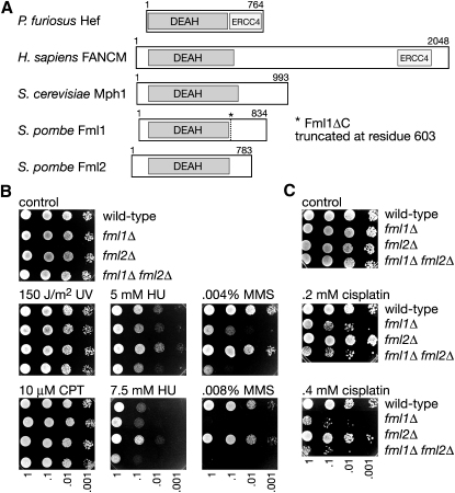
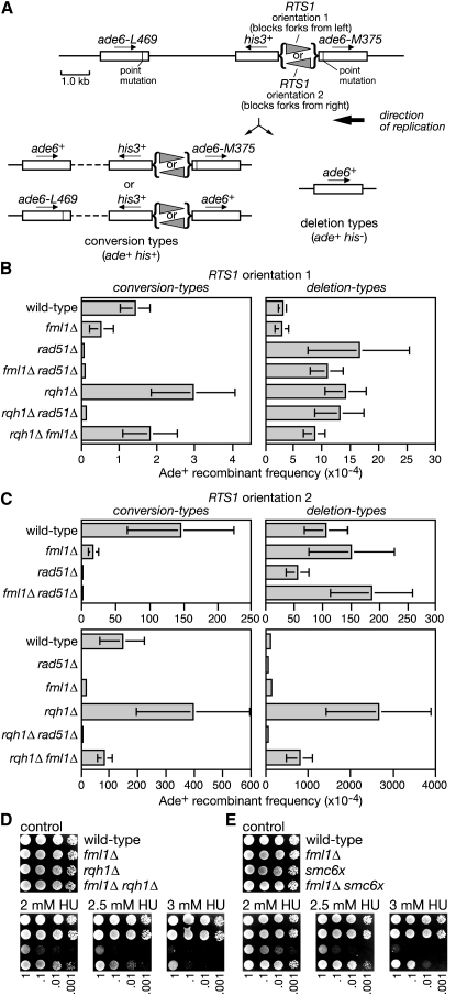
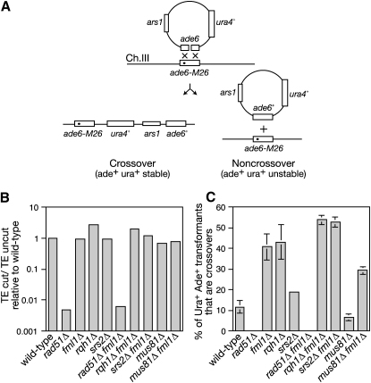
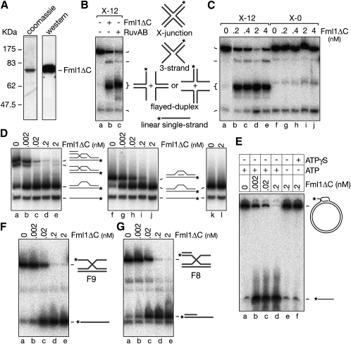
The Fanconi anemia (FA) core complex promotes the tolerance/repair of DNA damage at stalled replication forks by catalyzing the monoubiquitination of FANCD2 and FANCI. Intriguingly, the core complex component FANCM also catalyzes branch migration of model Holliday junctions and replication forks in vitro. Here we have characterized the ortholog of FANCM in fission yeast Fml1 in order to understand the physiological significance of this activity. We show that Fml1 has at least two roles in homologous recombination-it promotes Rad51-dependent gene conversion at stalled/blocked replication forks and limits crossing over during mitotic double-strand break repair. In vitro Fml1 catalyzes both replication fork reversal and D loop disruption, indicating possible mechanisms by which it can fulfill its pro- and antirecombinogenic roles.


Figures

Figure 1
Figure 1
Identification of a FANCM Ortholog in S. pombe (A) FANCM orthologs. DEAH helicase and ERCC4 nuclease domains are indicated. Note that the ERCC4 nuclease domain in FANCM is thought to be inactive. (B and C) Spot assays. The strains are MCW1221, MCW2080, MCW2078 and MCW2082.
Figure 2
Figure 2
Fml1 Promotes Spontaneous and RFB-Induced Recombination (A) Schematic showing the recombination substrate on chromosome 3 plus the two types of recombinant product. (B and C) Ade+ recombinant frequencies for strains (B) MCW1262, MCW3059, MCW1691, MCW3790, MCW1443, MCW2132, and MCW3444, and (C) MCW1433, MCW3061, MCW1692, MCW3794, MCW1447, MCW2130, and MCW3456. Error bars are the standard deviations about the mean. (D and E) Spot assays of strains (D) MCW1193, MCW2096, MCW1818, and MCW2487, and (E) MCW1221, MCW2080, MCW1712, and MCW3701.
Figure 3
Figure 3
Fml1 Limits Crossing Over during Mitotic DSB Repair (A) Schematic showing the repair of a double-strand gap in ade6 on plasmid pAN1 by homologous recombination with ade6-M26 on chromosome 3. The M26 mutation is indicated by the filled circle. See Figure S3 for a full description of the assay system. (B) Histogram showing the mean relative transformation efficiency (TE) of cut versus uncut plasmid in strains MCW1193, MCW2498, MCW2096, MCW1818, MCW3811, MCW2498, MCW2487, MCW2550, FO1192, and MCW2264. (C) Histogram showing the percentage of Ade+ recombinants that are crossovers in the same strains as in (B). Error bars are the standard deviations about the mean.
Figure 4
Figure 4
Catalysis of HJ Branch Migration and D Loop Dissociation by Fml1ΔC (A) Coomassie blue-stained SDS gel and immunoblot (probed with anti-polyhistidine) showing purified His-tagged Fml1ΔC. (B) PhosphorImage showing dissociation of X-12 by Fml1ΔC (2 nM) and RuvAB (40 nM RuvA and 630 nM RuvB). The schematic shows the various products of X-junction dissociation. Asterisks indicate 32P label at the DNA 5′ end. (C) Comparison of the dissociation of X-12 and X-0 by Fml1ΔC. (D) Dissociation of static D loops by Fml1ΔC. The substrates are D2 (lanes a–e), D7 (lanes f–j), and D8 (lanes k–l). The schematics show the DNA substrates and their various dissociation products, with the asterisks indicating the 32P label at the 5′ end of oligo 16. (E) Dissociation of a mobile D loop by Fml1ΔC. Reactions were incubated for 15 min at 37°C. (F and G) Dissociation of the part X-junctions F8 and F9 by Fml1ΔC.
Figure 5
Figure 5
Fml1ΔC Catalyzes Fork Reversal (A) Schematic of χ-DNA showing the position of restriction sites used to generate χKpn and χSma. The homologous core (gray lines) and size (in kb) of each duplex arm are indicated. (B) Comparison of χSma and χKpn cleavage by RuvC. (C) Schematic illustrating equilibrium between forms of χSma that are cleavable and non-cleavable by RuvC. (D) Stimulation of RuvC cleavage of χSma by increasing amounts of Fml1ΔC (0.01, 0.1 and 1 nM). (E) Quantification of the data in (D). (F) Rates of χSma and χKpn cleavage by RuvC in the presence and absence of Fml1ΔC. “C” and “F” denote RuvC and Fml1ΔC respectively. Values are means of two experiments (error bars are omitted for clarity). (G) Dependence on ATP hydrolysis for Fml1ΔC-stimulated cleavage of χSma by RuvC.
Figure 6
Figure 6
Lagging Strand Unwinding Catalyzed by Fml1ΔC (A–C) Unwinding assays showing Fml1ΔC's ability to dissociate fork substrates F2, F10 and F11. Schematics of the substrates and reaction products are shown with asterisks indicating the 32P label at the DNA 5′ end. (D and F) Time courses of F11 and F10 unwinding. Reactions (100 μl) contain Fml1ΔC (2 nM) and 32P labeled F11 or F10 (0.5 nM) as indicated. (E) Quantification of data in (D). (G) Quantification of data in (F).
Figure 7
Figure 7
Genetic Interactions with Other Junction Processing Enzymes and Models for Fml1's Roles in Replication Fork Processing and DSB Repair (A) Spot assay of strains MCW1221, MCW2080, MCW1238, and MCW2428. (B) Spot assay of strains MCW1221, MCW2080, MCW3781, and MCW3816. (C) Model for lesion bypass promoted by Fml1. (D) Model showing different pathways of DSB repair.

Similar articles

Cited by

References

    1. Ahn J.S., Osman F., Whitby M.C. Replication fork blockage by RTS1 at an ectopic site promotes recombination in fission yeast. EMBO J. 2005;24:2011–2023. - PMC - PubMed
    1. Ampatzidou E., Irmisch A., O'Connell M.J., Murray J.M. Smc5/6 is required for repair at collapsed replication forks. Mol. Cell. Biol. 2006;26:9387–9401. - PMC - PubMed
    1. Bachrati C.Z., Borts R.H., Hickson I.D. Mobile D-loops are a preferred substrate for the Bloom's syndrome helicase. Nucleic Acids Res. 2006;34:2269–2279. - PMC - PubMed
    1. Blastyak A., Pinter L., Unk I., Prakash L., Prakash S., Haracska L. Yeast Rad5 protein required for postreplication repair has a DNA helicase activity specific for replication fork regression. Mol. Cell. 2007;28:167–175. - PMC - PubMed
    1. Bugreev D.V., Mazina O.M., Mazin A.V. Rad54 protein promotes branch migration of Holliday junctions. Nature. 2006;442:590–593. - PubMed

Publication types

MeSH terms