

Review
. 2008 Oct 4;8 Spec No A(Spec Iss A):S33-42.
doi: 10.1102/1470-7330.2008.9007.
Staging esophageal cancer
Affiliations
- PMID: 18852079
- PMCID: PMC2582495
- DOI: 10.1102/1470-7330.2008.9007
Item in Clipboard
Review
Staging esophageal cancer
Cancer Imaging.
.
Abstract
Accurate staging of disease is necessary in patients with newly diagnosed esophageal cancer in order to prompt appropriate curative or palliative therapy. Computed tomography (CT) may be used to evaluate for local spread into adjacent structures (T4 disease) and to diagnose distant metastases (M1). Endoscopic ultrasonography (EUS) is the modality of choice for distinguishing T1 tumors from higher stage lesions and for detecting and sampling regional lymph nodes (N1 disease). Positron emission tomography (PET) scanning is most helpful for detecting previously occult distant metastases. Optimal staging generally requires a multimodality approach.
Figures
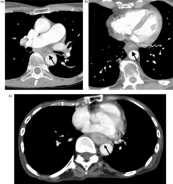
Three patients with broad loss of the fat plane between the esophagus and the aorta (arrows). At surgery, there was no aortic invasion in (A) (squamous cell carcinoma) or (B) (adenocarcinoma), however aortic invasion was present in (C) (squamous cell carcinoma).
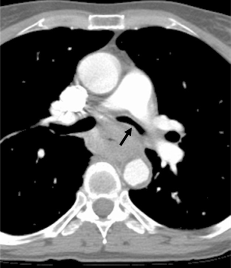
Esophageal adenocarcinoma causing narrowing of the left mainstem bronchus at CT (arrow). Airway invasion was confirmed bronchoscopically.
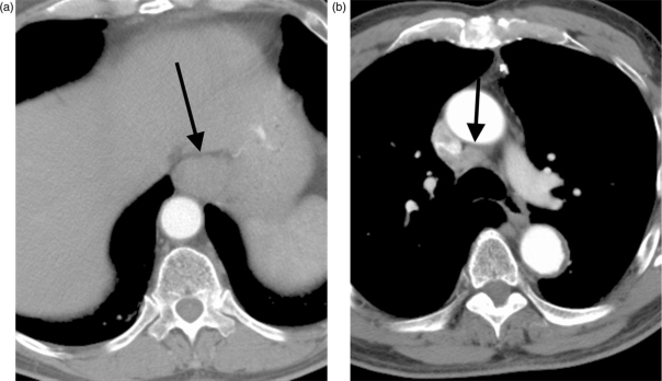
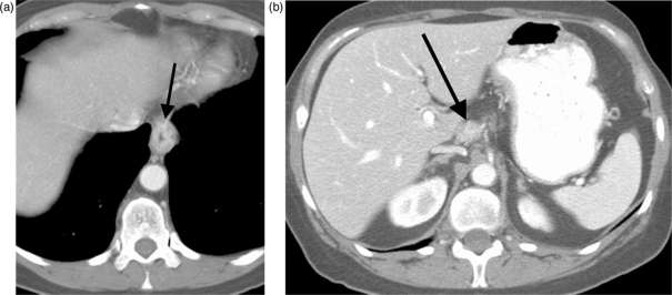
Distal esophageal adenocarcinoma (arrow, A) and right paratracheal lymph node enlargement (arrow, B), suggesting regional nodal metastatic disease. Lymph node biopsy, however, revealed no malignancy.
FDG avid distal esophageal adenocarcinoma (arrow) on PET-CT scanning (A–C). Regional nodal (N1) disease (found at surgery) was not diagnosed preoperatively at PET, probably because uptake in the primary tumor obscured the abnormal, adjacent lymph nodes.
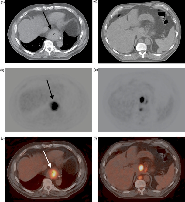
FDG avid distal esophageal adenocarcinoma (arrow, A–C) on PET-CT scanning. The CT image helps to localize nearby foci of FDG avidity to abnormal lymph nodes in the gastrohepatic ligament and retrocrural region (D–F).
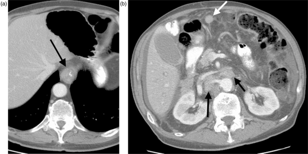
Primary distal esophageal adenocarcinoma (arrow, A) with extensive distant metastatic disease to sites including retroperitoneal lymph nodes (black arrows, B) and peritoneum (white arrow, B). The findings represent M1b disease.
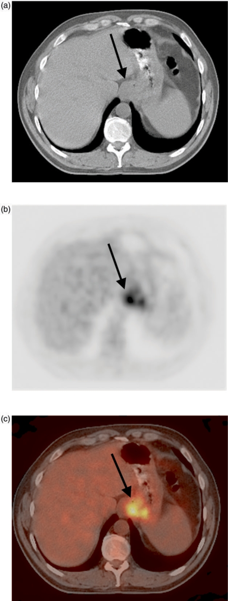
Distal esophageal adenocarcinoma (arrow, A) with metastasis in a celiac axis lymph node (arrow, B), constituting M1a disease.
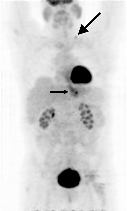
Projection PET image shows an FDG-avid distal esophageal adenocarcinoma (small arrow) and a previously occult distant metastasis within a left supraclavicular lymph node (large arrow), constituting M1b disease.
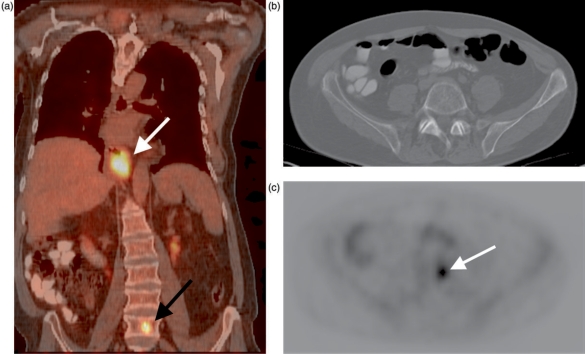
Fused PET-CT image reveals an FDG-avid distal esophageal carcinoma (white arrow, A) and a previously occult distant vertebral body metastasis (black arrow, A). The spine appears normal on an axial CT image at the level of the metastasis (B); axial PET image at the same level shows the lesion (arrow, C).
References
-
- Bogot N, Quint L. CT in esophageal cancer. In: Rankin SC, editor. Carcinoma of the esophagus. Cambridge: Cambridge University Press; 2008. pp. p. 62–84.
-
- New York: Springer-Verlag; 2002. AJCC cancer staging handbook.
-
- Patti MG, Gantert W, Way LW. Surgery of the esophagus. Anatomy and physiology. Surg Clin North Am. 1997;77:959–70. - PubMed
-
- Riquet M, Saab M, Le Pimpec Barthes F, Hidden G. Lymphatic drainage of the esophagus in the adult. Surg Radiol Anat. 1993;15:209–11. - PubMed
-
- SEER NCI. 2008. Cancer of the esophagus. http://seer.cancer.gov/statfacts/html/esoph.html.
Publication types
MeSH terms
LinkOut - more resources
Full Text Sources
Medical