

Hepatocellular carcinoma: development and early detection
- PMID: 18852086
- PMCID: PMC2582507
- DOI: 10.1102/1470-7330.2008.9019
Hepatocellular carcinoma: development and early detection
Abstract
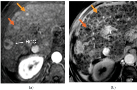
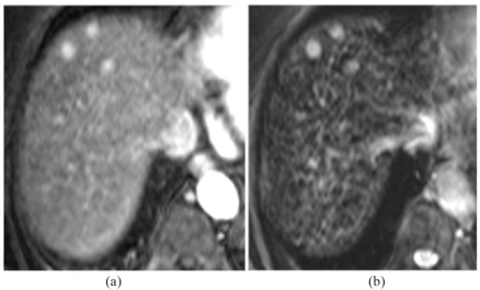
Our eventual aim is to predict, using non-invasive imaging techniques, the biological behaviour of individual cirrhotic nodules. We are some distance away from this, so our current objective is to define imaging features which predict the histologic findings. This short review summarises the current capabilities and limitations of non-invasive imaging in detecting small hepatocellular carcinomas (HCCs) in cirrhosis. Extracellular contrast media used with ultrasound (US), computed tomography (CT) or magnetic resonance imaging (MRI) can characterise nodules according to the predominance of arterial or portal inflow, and most HCCs will be recognised by their arterial hypervascularity. Adding intracellular (liver-specific) MRI contrast agents provides a significant improvement in early detection and in specificity for HCC. Nodules can be classified on dual contrast MRI as clearly malignant, clearly benign, or borderline (needing careful surveillance). Future imaging research needs to establish the histology of small hypervascular nodules, the evolution of hypervascular nodules and of dysplastic nodules, and to seek imaging features which predict microvascular invasion. Currently, cirrhotic patients with either suspicious nodules on screening US or rising AFP should have cross-sectional imaging with multi-phase CT or preferably MRI. Dual-contrast MRI with liver-specific agents should be used to improve diagnostic specificity for small lesions. Borderline nodules should be followed at agreed intervals using the same imaging technique each time. Pre-operative staging in surgical candidates should include CT of thorax, abdomen and pelvis and bone scintigraphy.
Figures
References
-
- Ward J, Guthrie JA, Scott DJ, et al. Hepatocellular carcinoma in the cirrhotic liver: double-contrast MR imaging for diagnosis. Radiology. 2000;216:154–62. - PubMed
-
- Jeong YY, Mitchell DG, Kamishima T. Small (<20 mm) enhancing hepatic nodules seen on arterial phase MR imaging of the cirrhotic liver: clinical implications. Am J Roentgenol. 2002;178:1327–34. - PubMed
-
- Burrel M, Llovet JM, Avuso C, et al. MRI angiography is superior to helical CT for detection of HCC prior to liver transplantation: an explant correlation. Hepatology. 2003;38:1034–42. - PubMed
-
- Bhartia B, Ward J, Guthrie JA, Robinson PJ. Hepatocellular carcinoma in cirrhotic livers: double-contrast thin-section MR imaging with pathologic correlation of explanted tissue. Am J Roentgenol. 2003;180:577–84. - PubMed
-
- Kwak HS, Lee JM, Kim CS. Preoperative detection of hepatocellular carcinoma: comparison of combined contrast-enhanced MR imaging and combined CT during arterial portography and CT hepatic arteriography. Eur Radiol. 2004;14:447–57. - PubMed
Publication types
MeSH terms
LinkOut - more resources
Full Text Sources
Medical