. 2008 Oct;7(10):3169-75.
doi: 10.1158/1535-7163.MCT-08-0314.

Stat3 contributes to resistance toward BCR-ABL inhibitors in a bone marrow microenvironment model of drug resistance

Affiliations

Stat3 contributes to resistance toward BCR-ABL inhibitors in a bone marrow microenvironment model of drug resistance

Nadine N Bewry et al. Mol Cancer Ther. 2008 Oct.

Abstract

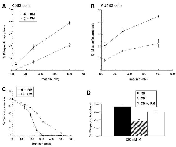
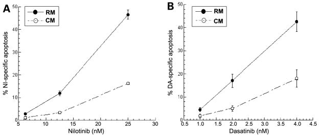
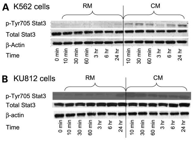
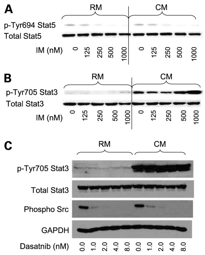
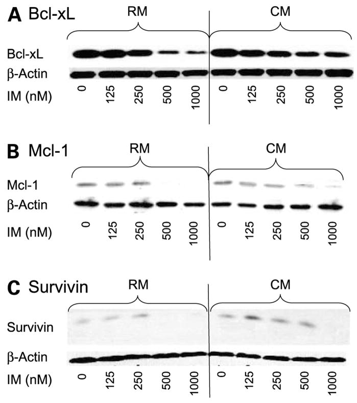
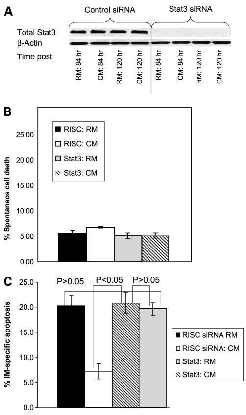
Imatinib mesylate is a potent, molecularly targeted therapy against the oncogenic tyrosine kinase BCR-ABL. Although imatinib mesylate has considerable efficacy against chronic myeloid leukemia (CML), advanced-stage CML patients frequently become refractory to this agent. The bone marrow is the predominant microenvironment of CML and is a rich source of both soluble factors and extracellular matrices, which may influence drug response. To address the influence of the bone marrow microenvironment on imatinib mesylate sensitivity, we used an in vitro bone marrow stroma model. Our data show culturing K562 cells, in bone marrow stroma-derived conditioned medium (CM), is sufficient to cause resistance to BCR-ABL inhibitors. Drug resistance correlated with increased pTyrStat3, whereas no increases in pTyrStat5 was noted. Moreover, resistance was associated with increased levels of the Stat3 target genes Bcl-xl, Mcl-1, and survivin. Finally, reducing Stat3 levels with small interfering RNA sensitized K562 cells cultured in CM to imatinib mesylate-induced cell death. Importantly, Stat3 dependency was specific for cells grown in CM, as reducing Stat3 levels in regular growth conditions had no effect on imatinib mesylate sensitivity. Together, these data support a novel mechanism of BCR-ABL-independent imatinib mesylate resistance and provides preclinical rationale for using Stat3-inhibitors to increase the efficacy of imatinib mesylate within the context of the bone marrow microenvironment.


Conflict of interest statement

Disclosure of Potential Conflicts of Interest

No potential conflicts of interest were disclosed.

Figures

Figure 1
Figure 1
HS-5-derived CM protects K562 and KU182 CML cells from death induced by imatinib mesylate. K562 (A) or KU182 (B) cells were cultured in either RM or CM for 3 h and before the addition of various concentrations of imatinib mesylate or 0.1% DMSO for 36 h. Cell death was measured using Annexin V apoptosis assay followed by fluorescence-activated cell sorting analysis. % Drug-specific apoptosis was calculated by subtracting the background cell death in control DMSO-treated cells from drug treated cell death. A, CM significantly protects K562 cells from imatinib mesylate-mediated cell death (P < 0.05, analysis of covariance). B, CM significantly protects KU182 cells from imatinib mesylate-mediated cell death (P < 0.05, analysis of covariance). C, CM increases the clonogenic survival of K562 CML cells treated with imatinib mesylate. K562 cells were cultured in RM or CM and treated with increasing concentrations of imatinib mesylate as depicted. After 6 h incubation at 37°C, 5% CO2, cells were cultured in 0.3% agar made of RM or CM and containing their respective imatinib mesylate concentrations. Cells were allowed to incubate at 37°C, 5% CO2 for 10 d. Cell colonies (>50 cells) were counted. Representative figure in triplicate (n = 3 independent experiments). D, removal of CM partially reverses resistance to imatinib mesylate. K562 cells were grown in either CM or RM for 3 h. Following 3 h, appropriate samples were removed from CM and placed in RM for an additional 3 h. Cells were treated with 500 nmol/L imatinib mesylate for 36 h and apoptosis was measured by Annexin V positivity. Representative figure in triplicates.
Figure 2
Figure 2
Cells were allowed to incubate for either 48 h (nilotinitib) or 24 h (dasatinib). CM significantly protects K562 cells from (A) nilotinib-mediated cell death (P < 0.05, analysis of covariance) and (B) dasatinib-mediated cell death (P < 0.05, analysis of covariance). Representative graph in triplicates (n = 2 independent experiments).
Figure 3
Figure 3
Stat3 pTyr705 is increased in CM. A, K652 cells were cultured in RM or CM for the time points indicated. Cells were collected, lysed, and analyzed for either pTyr705 or total Stat3 via Western blotting. β-Actin was used as a loading control. B, KU812 cells were cultured in RM or CM for the time points indicated. Cells were collected, lysed, and analyzed for either pTyr705 or total Stat3 via Western blotting. β-Actin was used as a loading control. Representative figure (n = 3 independent experiments).
Figure 4
Figure 4
A, K562 cells grown in CM show equal levels of pTyrStat5 and equal inhibition of pTyrStat5 levels following 48 h of imatinib mesylate treatment compared with cells treated in RM. B, treating K562 cells with imatinib mesylate for 48 h did not reduce the levels of pStat3 when K562 cells were cultured in CM. C, treating K562 cells with dasatinib inhibited levels of pSrc family members but did not attenuate the levels of pStat3 when K562 cells were cultured in CM. Representative figure (n = 3 independent experiments).
Figure 5
Figure 5
Effects of CM on Stat3 downstream targets in CM. K562 CML cells were treated with imatinib mesylate in a dose-dependent manner for 48 h. Western blot analysis was done with specific antibodies to Bcl-xl, Mcl-1, and survivin as described in Materials and Methods. A, Bcl-xl; B, Mcl-1; C, survivin. β-Actin was used as a loading control. Representative figure (n = 3 independent experiments).
Figure 6
Figure 6
Reducing Stat3 levels with siRNA reverses resistance to imatinib mesylate when K562 cells are grown in CM. K562 cells were cultured in either RM or CM and treated with either siRNA to Stat3 or RISC control siRNA. A, Stat3 knockdown was confirmed using Western blotting and Stat3 was noted to be maximally decreased at 84 h and remained reduced for at least 120 h (n = 5 independent experiments). B, reducing Stat3 levels was not sufficient to cause cell death in cells cultured in RM or CM. Mean ± SD of 5 independent experiments. C, reducing Stat3 levels enhances sensitivity to imatinib mesylate when K562 cells are cultured in CM. Apoptosis of K562 cells cultured in RM or CM, treated with either Stat3 siRNA or RISC control siRNA, in the presence or absence of 250 nmol/L imatinib mesylate was done using the Annexin V detection of apoptotic cells and fluorescence-activated cell sorting analysis. % Specific apoptosis was calculated by subtracting background cell death from imatinib mesylate-mediated cell death. Representative figure in triplicates (n = 5 independent experiments).

References

    1. Bartram CR, de Klein A, Hagemeijer A, et al. Translocation of c-ab1 oncogene correlates with the presence of a Philadelphia chromosome in chronic myelocytic leukaemia. Nature. 1983;306:277–80. - PubMed
    1. Groffen J, Stephenson JR, Heisterkamp N, de Klein A, Bartram CR, Grosveld G. Philadelphia chromosomal breakpoints are clustered within a limited region, bcr, on chromosome 22. Cell. 1984;36:93–9. - PubMed
    1. Lugo TG, Pendergast AM, Muller AJ, Witte ON. Tyrosine kinase activity and transformation potency of bcr-abl oncogene products. Science. 1990;247:1079–82. - PubMed
    1. Buchdunger E, Zimmermann J, Mett H, et al. Inhibition of the Abl protein-tyrosine kinase in vitro and in vivo by a 2-phenylaminopyrimidine derivative. Cancer Res. 1996;56:100–4. - PubMed
    1. Druker BJ, Guilhot F, O’Brien SG, et al. Five-year follow-up of patients receiving imatinib for chronic myeloid leukemia. N Engl J Med. 2006;355:2408–17. - PubMed

Publication types

MeSH terms