. 2008 Dec;76(12):5553-64.
doi: 10.1128/IAI.00885-08. Epub 2008 Oct 13.

Vaccine-induced cellular immune responses differ from innate responses in susceptible and resistant strains of mice infected with Coccidioides posadasii

Affiliations

Vaccine-induced cellular immune responses differ from innate responses in susceptible and resistant strains of mice infected with Coccidioides posadasii

Lisa F Shubitz et al. Infect Immun. 2008 Dec.

Abstract

Susceptibility to Coccidioides spp. varies widely in humans and other mammals and also among individuals within a species. Among strains of mice with various susceptibilities, immunohistopathology revealed that C57BL/6 mice were highly susceptible to the disease following intranasal infection, DBA/2n mice were intermediate, and Swiss-Webster mice were innately resistant. Resistant Swiss-Webster mice developed prominent perivascular/peribronchiolar lymphocytic cuffing and well-formed granulomas with few fungal elements and debris in the necrotic center, surrounded by a mantle of macrophages, lymphocytes, and fibrocytes. Susceptible C57BL/6 mice became moribund between 14 and 18 days postinfection, with overwhelming numbers of neutrophils and spherules and very few T cells, the drastic reduction of which was associated with failure and death, while intermediate DBA/2n mice controlled the fungal burden but demonstrated progressive lung inflammation with prominent suppuration, and they deteriorated clinically. Vaccinated C57BL/6 mice had an early and robust lymphocyte response, which included significantly higher Mac2(+), CD3(+), and CD4(+) cell scores on day 18 than those of innately resistant SW mice and DBA/2n mice; they also had prominent perivascular/peribronchiolar lymphocytic infiltrates not present in their unvaccinated counterparts, and they appeared to be resolving lesions by day 56 compared to the other two strains, based on significantly lower disease scores and observably smaller and fewer lesions with few spherules and neutrophils.


Figures

FIG. 1.
FIG. 1.
Numbers of CFU in the right lung (Rt lung) of B6, D2, and SW mice after intranasal infection with Coccidioides posadasii. (A) B6 and D2 (n = 4) mice received 50 spores; numbers of CFU were significantly greater in B6 mice on days 12 and 18 (P < 0.05). (B) B6 and SW mice (n = 8) received 65 spores (target dose = 50 spores); numbers of CFU were significantly different on all days together (P < 0.05).
FIG. 2.
FIG. 2.
Lung fungal burdens in unvaccinated and vaccinated B6, D2, and SW mice (n = 8) at days 12, 18, and 56 postinfection. SW mice have significantly fewer fungal CFU than B6 mice (P < 0.001) and D2 mice (P = 0.001) at all time points. Vaccinated B6 mice have significantly fewer fungal CFU than unvaccinated B6 mice (P < 0.001), and there is a statistical difference between all vaccinated mice and unvaccinated mice, but strain-specific analysis reveals that all the significance comes from the B6 mice.
FIG. 3.
FIG. 3.
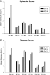
Mean spherule score from slides stained with the Coccidioides-specific anti-Ag2/PRA antibody (A) or gross disease score at necropsy (B) by day, vaccination status, and mouse strain. These parallel the quantitative results shown in Fig. 2 quite well with the exception of the notably high disease score in D2 mice compared to their fungal burdens on days 18 and 56. Examination of slides revealed that D2 mice have severe inflammation, though they appear to be controlling the fungal burden.
FIG. 4.
FIG. 4.
Lesions at day 6 postinfection. (A) Neutrophils are the most abundant cells in the microabscess, with macrophages (arrowheads) and small lymphocytes seen occasionally. Magnification, ×400; hematoxylin-and-eosin stain. The microabscesses in SW and B6 mice (B) have a distinct border with the normal tissue, while lesions in D2 mice (C) exhibit hemorrhage and extensive edema. Magnification, ×40; hematoxylin-and-eosin stain.
FIG. 5.
FIG. 5.
(A to D) Representative sections from day 18 postinfection. Unvaccinated B6 mice have a myriad of spherules and neutrophils with minimal perivascular cuffing (A); D2 mice have fewer obvious spherules than B6 mice, but the lesion is extensive, with hemorrhage and edema (B); both SW mice (C) and vaccinated B6 mice (D) have smaller lesions with distinct borders, few organisms, and significant PV/PB cuffing. (E to H) Day-18-postinfection sections stained with Coccidioides-specific antibody highlight the enormity of spherule burden in unvaccinated B6 mice (E), the extent of lesions despite a relatively controlled spherule burden in D2 mice (F), and the low fungal numbers with controlled lesion size in SW mice (G) and vaccinated B6 mice (H). (I to K) On day 56 postinfection, inflammation is extensive and disorganized in D2 mice (I), with multifocal neutrophilic islands surrounded by poorly organized macrophages (I, inset); SW mice (J) and vaccinated B6 mice (K) have only one or a few small, contained lesions per section and thick PV/PB infiltrates. SW mice also demonstrate lymphoid aggregates at the lesion margins. Magnification, ×40. Panels A to D and I to K show hematoxylin-and-eosin staining; panels E to H show anti-Ag2/PRA antibody; I inset shows Mac2 antibody.
FIG. 6.
FIG. 6.
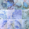
Representative images of sections stained for Mac2, CD3, and CD4 on day 18 postinfection. (A to D) B6 mice (A) have undergone regression of macrophages compared to results on day 6 (A, inset), while D2 mice (B) have expanding lesions with many macrophages. SW mice (C) and B6 mice (D) are similar in having relatively small lesions with a band of macrophages surrounding a necrotic center, though the scores for Mac2 on day 18 were statistically higher for B6 mice than for SW mice (Fig. 6). (E to H) Unvaccinated B6 mice are shown at magnification ×200 to highlight the few CD3 cells present by day 18. CD3+ cells in D2 mice (F) are most prominent in the PV/PB infiltrates and scattered in the lesion margins, while SW mice (G) and vaccinated B6 mice (H) have abundant CD3+ in the lesions. (I to L) CD4+ cells parallel the CD3+ cells in location, though fewer in number. Note the composition of the marginal lymphoid aggregate from an SW mouse (K inset) and the abundance of them in the vaccinated B6 mouse (L) compared to the SW mouse (P = 0.003). Magnification for panels A to D, ×40; magnification for panel E, ×200; magnification for panels F to L, ×100.
FIG. 7.
FIG. 7.
Mean scores for CD3, CD22, and Mac2 in vaccinated and unvaccinated B6 and SW mice on days 12 and 18 (n = 8 for each bar). CD3+, CD22+, and Mac2+ cells are increased in vaccinated B6 mice compared to those in SW mice, with CD3+ and Mac2+ cells significantly increased on day 18 (P < 0.001 and P = 0.02, respectively). In unvaccinated B6 mice, both lymphocyte populations are smaller than those in SW mice and CD3+ cells are highly significantly reduced on day 18 (P = 0.006).

References

    1. Abuodeh, R. O., L. F. Shubitz, E. Siegel, S. Snyder, T. Peng, K. I. Orsborn, E. Brummer, D. A. Stevens, and J. N. Galgiani. 1999. Resistance to Coccidioides immitis in mice after immunization with recombinant protein or a DNA vaccine of a proline-rich antigen. Infect. Immun. 672935-2940. - PMC - PubMed
    1. Baldridge, J. B., and R. T. Crane. 1999. Monophosphoryl lipid A (MPL) formulations for the next generation of vaccines. Methods 19103-107. - PubMed
    1. Baldwin, S. L., C. D'Souza, A. D. Roberts, B. P. Kelly, A. A. Frank, M. A. Lui, J. B. Ulmer, K. Huygen, D. M. McMurray, and I. M. Orme. 1998. Evaluation of new vaccines in the mouse and guinea pig model of tuberculosis. Infect. Immun. 662951-2959. - PMC - PubMed
    1. Beaman, L., D. Pappagianis, and E. Benjamini. 1977. Significance of T cells in resistance to experimental murine coccidioidomycosis. Infect. Immun. 17580-585. - PMC - PubMed
    1. Beaman, L., D. Pappagianis, and E. Benjamini. 1979. Mechanisms of resistance to infection with Coccidioides immitis in mice. Infect. Immun. 23681-685. - PMC - PubMed

Publication types

Substances