

Infected-host-cell repertoire and cellular response in the lung following inhalation of Francisella tularensis Schu S4, LVS, or U112
- PMID: 18852251
- PMCID: PMC2583552
- DOI: 10.1128/IAI.01176-08
Infected-host-cell repertoire and cellular response in the lung following inhalation of Francisella tularensis Schu S4, LVS, or U112
Abstract
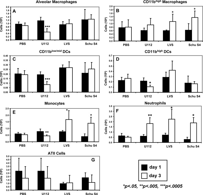
Francisella tularensis causes systemic disease in humans and other mammals, with high morbidity and mortality associated with inhalation-acquired infection. F. tularensis is a facultative intracellular pathogen, but the scope and significance of cell types infected during disease is unknown. Using flow cytometry, we identified and quantified infected-cell types and assessed the impact of infection on cell populations following inhalation of F. tularensis strains U112, LVS, and Schu S4. Initially, alveolar macrophages comprised over 70% of Schu S4- and LVS-infected cells, whereas approximately 51% and 27% of U112-infected cells were alveolar macrophages and neutrophils, respectively. After 3 days, roughly half of Schu S4- and LVS- and nearly 80% of U112-infected cells were neutrophils. All strains infected CD11b(high) macrophages, dendritic cells, monocytes, and alveolar type II cells throughout infection. Macrophage, monocyte, and dendritic-cell populations were reduced during U112 infection but not Schu S4 or LVS infection. These results demonstrate directly that F. tularensis is a promiscuous intracellular pathogen in the lung that invades and replicates within cell types ranging from migratory immune cells to structural tissue cells. However, the proportions of cell types infected and the cellular immune response evoked by the human pathogenic strain Schu S4 differ from those of the human avirulent U112.
Figures
Similar articles
-
Interactions of Francisella tularensis with Alveolar Type II Epithelial Cells and the Murine Respiratory Epithelium.PLoS One. 2015 May 26;10(5):e0127458. doi: 10.1371/journal.pone.0127458. eCollection 2015. PLoS One. 2015. PMID: 26010977 Free PMC article.
-
Long-Term Survival of Virulent Tularemia Pathogens outside a Host in Conditions That Mimic Natural Aquatic Environments.Appl Environ Microbiol. 2021 Feb 26;87(6):e02713-20. doi: 10.1128/AEM.02713-20. Print 2021 Feb 26. Appl Environ Microbiol. 2021. PMID: 33397692 Free PMC article.
-
Monophosphoryl Lipid A Enhances Efficacy of a Francisella tularensis LVS-Catanionic Nanoparticle Subunit Vaccine against F. tularensis Schu S4 Challenge by Augmenting both Humoral and Cellular Immunity.Clin Vaccine Immunol. 2017 Mar 6;24(3):e00574-16. doi: 10.1128/CVI.00574-16. Print 2017 Mar. Clin Vaccine Immunol. 2017. PMID: 28077440 Free PMC article.
-
Host-pathogen interactions and immune evasion strategies in Francisella tularensis pathogenicity.Infect Drug Resist. 2014 Sep 18;7:239-51. doi: 10.2147/IDR.S53700. eCollection 2014. Infect Drug Resist. 2014. PMID: 25258544 Free PMC article. Review.
-
Multifaceted effects of Francisella tularensis on human neutrophil function and lifespan.Immunol Rev. 2016 Sep;273(1):266-81. doi: 10.1111/imr.12445. Immunol Rev. 2016. PMID: 27558340 Free PMC article. Review.
Cited by
-
Current status of vaccine development for tularemia preparedness.Clin Exp Vaccine Res. 2013 Jan;2(1):34-9. doi: 10.7774/cevr.2013.2.1.34. Epub 2013 Jan 15. Clin Exp Vaccine Res. 2013. PMID: 23596588 Free PMC article.
-
Stringent response governs the oxidative stress resistance and virulence of Francisella tularensis.PLoS One. 2019 Oct 24;14(10):e0224094. doi: 10.1371/journal.pone.0224094. eCollection 2019. PLoS One. 2019. PMID: 31648246 Free PMC article.
-
Francisella tularensis harvests nutrients derived via ATG5-independent autophagy to support intracellular growth.PLoS Pathog. 2013 Aug;9(8):e1003562. doi: 10.1371/journal.ppat.1003562. Epub 2013 Aug 15. PLoS Pathog. 2013. PMID: 23966861 Free PMC article.
-
Identification of early interactions between Francisella and the host.Infect Immun. 2014 Jun;82(6):2504-10. doi: 10.1128/IAI.01654-13. Epub 2014 Mar 31. Infect Immun. 2014. PMID: 24686053 Free PMC article.
-
Francisella tularensis Confronts the Complement System.Front Cell Infect Microbiol. 2017 Dec 19;7:523. doi: 10.3389/fcimb.2017.00523. eCollection 2017. Front Cell Infect Microbiol. 2017. PMID: 29312899 Free PMC article.
References
-
- Allen, L. A., and R. L. McCaffrey. 2007. To activate or not to activate: distinct strategies used by Helicobacter pylori and Francisella tularensis to modulate the NADPH oxidase and survive in human neutrophils. Immunol. Rev. 219103-117. - PubMed
-
- Bosio, C. M., H. Bielefeldt-Ohmann, and J. T. Belisle. 2007. Active suppression of the pulmonary immune response by Francisella tularensis Schu4. J. Immunol. 1784538-4547. - PubMed
-
- Bosio, C. M., and S. W. Dow. 2005. Francisella tularensis induces aberrant activation of pulmonary dendritic cells. J. Immunol. 1756792-6801. - PubMed
Publication types
MeSH terms
Grants and funding
LinkOut - more resources
Full Text Sources
Other Literature Sources
Research Materials
