. 2008 Dec 5;283(49):33816-25.
doi: 10.1074/jbc.M807428200. Epub 2008 Oct 13.

Abnormal metabolism of glycogen phosphate as a cause for Lafora disease

Affiliations

Abnormal metabolism of glycogen phosphate as a cause for Lafora disease

Vincent S Tagliabracci et al. J Biol Chem. .

Abstract

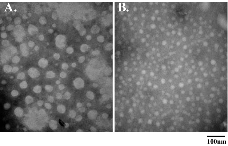
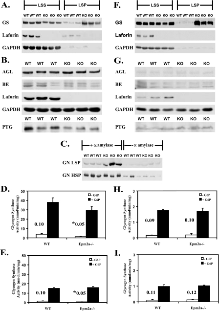
Lafora disease is a progressive myoclonus epilepsy with onset in the teenage years followed by neurodegeneration and death within 10 years. A characteristic is the widespread formation of poorly branched, insoluble glycogen-like polymers (polyglucosan) known as Lafora bodies, which accumulate in neurons, muscle, liver, and other tissues. Approximately half of the cases of Lafora disease result from mutations in the EPM2A gene, which encodes laforin, a member of the dual specificity protein phosphatase family that is able to release the small amount of covalent phosphate normally present in glycogen. In studies of Epm2a(-/-) mice that lack laforin, we observed a progressive change in the properties and structure of glycogen that paralleled the formation of Lafora bodies. At three months, glycogen metabolism remained essentially normal, even though the phosphorylation of glycogen has increased 4-fold and causes altered physical properties of the polysaccharide. By 9 months, the glycogen has overaccumulated by 3-fold, has become somewhat more phosphorylated, but, more notably, is now poorly branched, is insoluble in water, and has acquired an abnormal morphology visible by electron microscopy. These glycogen molecules have a tendency to aggregate and can be recovered in the pellet after low speed centrifugation of tissue extracts. The aggregation requires the phosphorylation of glycogen. The aggregrated glycogen sequesters glycogen synthase but not other glycogen metabolizing enzymes. We propose that laforin functions to suppress excessive glycogen phosphorylation and is an essential component of the metabolism of normally structured glycogen.


Figures

FIGURE 1.
FIGURE 1.
Glycogen and glycogen phosphate levels increase with age in the absence of laforin. A, glycogen phosphate levels in muscle glycogen from wild type (WT) and Epm2a–/– mice at 3 months (open bars) and 9–12 months (filled bars). *, p < 0.001 versus wild type; #, p < 0.01 versus 3-month-old Epm2a–/– (n = 7–8). B, skeletal muscle glycogen content levels in wild type and Epm2a–/– mice. *, p < 0.001 versus WT and 3-month-old Epm2a–/– (n = 11–15). C, brain glycogen content in WT and Epm2a–/– mice. *, p = 0.0001 versus WT; #, p < 0.002 versus 3-month-old Epm2a–/– (n = 3–4).
FIGURE 2.
FIGURE 2.
Age-dependent changes in chemical and physical properties of glycogen in Epm2a–/– mice. A, iodine spectra of skeletal muscle glycogen (50–75 μg/ml) purified from wild type (WT) and Epm2a–/– mice. Amylopectin is shown for reference. B, quantitation of A.*, p < 0.002 versus wild type; #, p < 0.0001 versus 3-month-old Epm2a–/– (n = 3–8). C, skeletal muscle glycogen (5 mg/ml) purified from a 9-month-old Epm2a–/– mice that was treated with catalytically inactive laforin (C266S) (tube i) or active laforin (tube ii), and brought to 66% (v/v) ethanol. Tube iii, addition of LiCl to tube i. Tube iv, addition of LiCl to tube ii. Tube v, addition of LiCl to a 66% ethanol solution without glycogen.
FIGURE 3.
FIGURE 3.
Age-dependent changes in glycogen structure in Epm2a–/– mice. Electron micrographs of skeletal muscle glycogen purified from: 3-month-old wild type (WT)(A); 9–12-month-old wild type (B); 3-month-old Epm2a–/– (C); 9–12-month-old Epm2a–/– (D); and size distribution of skeletal muscle glycogen particles purified from wild type and Epm2a–/– mice (E).
FIGURE 4.
FIGURE 4.
Effect of phosphate removal on skeletal muscle glycogen from 9–12-month-old Epm2a–/– mice. Electron micrographs of glycogen treated with the catalytically inactive laforin (C266S) (A) or active wild type laforin (B).
FIGURE 5.
FIGURE 5.
Fractionation of glycogen from 9–12-month-old Epm2a–/– mice. Total skeletal muscle or brain extracts from 9–12-month-old wild type (WT) and Epm2a–/– mice were subjected to low speed centrifugation to generate supernatant (LSS) and pellet (LSP). A, percentage of total muscle glycogen, in the LSP. *, p < 0.05 (n = 3). B, percentage of total brain glycogen in the LSP. *, p < 0.005 (n = 3). C, electron micrograph of glycogen purified from the LSS of a muscle extract. D, electron micrograph of glycogen purified from the LSP of a muscle extract. E, skeletal muscle glycogen phosphate levels in LSS and the LSP. #, p = 0.002 (n = 6). F, iodine spectra of muscle glycogen (50–75 μg/ml) (purified from the LSS and the LSP; n = 6). Amylopectin is shown for reference.
FIGURE 6.
FIGURE 6.
Analysis of glycogen metabolizing enzymes and related proteins in old Epm2a–/– mice. Muscle or brain extracts from 9–12-month-old wild type (WT) and Epm2a–/– mice were analyzed. A, GS protein levels in the LSS and the LSP of muscle extracts. B, glycogen debranching enzyme (AGL) and branching enzyme (BE) protein levels in muscle extracts, and muscle PTG protein levels in the high speed centrifugation glycogen pellet. C, glycogenin protein levels in the LSP and the high speed pellets (HSP) of muscle treated or not with α-amylase. D, GS activity in LSS from muscle in the absence (white bars) or presence (black bars) of G6P. The corresponding activity ratio is shown. *, p < 0.05 (n = 3). E, GS activity in the LSP from muscle in the absence or presence of G6P. The corresponding activity ratio is shown. *, p < 0.05 (n = 3). F, GS protein levels in the LSS and the LSP of brain extracts. G, AGL and branching enzyme protein levels in brain extracts, and PTG protein levels in the high speed centrifugation glycogen pellet of brain extracts. H, GS activity in the LSS from brain. The activity ratio is shown (n = 3). I, GS activity in the LSP from brain. The activity ratio is shown (n = 3). KO, knock-out.
FIGURE 7.
FIGURE 7.
Possible roles for laforin in malin in the formation of polyglucosan. We propose that laforin is a physiological glycogen phosphatase whose impairment leads to the structural abnormalities in glycogen described in this work and to Lafora body formation. The role of malin remains a matter of active investigation. Malin could act together with or upstream of laforin in the removal of phosphate from glycogen (a). Malin could function independently of laforin to reduce glycogen phosphorylation, such as by down-regulating the enzyme(s) responsible for glycogen phosphorylation (b). Alternatively, malin could suppress polyglucosan formation independently of glycogen phosphorylation by controlling glycogen metabolic enzymes (c), as has been proposed.

Similar articles

Cited by

References

    1. Roach, P. J. (2002) Curr. Mol. Med. 2 101–120 - PubMed
    1. Ball, S., Guan, H. P., James, M., Myers, A., Keeling, P., Mouille, G., Buleon, A., Colonna, P., and Preiss, J. (1996) Cell 86 349–352 - PubMed
    1. Gunja-Smith, Z., Marshall, J. J., Mercier, C., Smith, E. E., and Whelan, W. J. (1970) FEBS Lett. 12 101–104 - PubMed
    1. Shearer, J., and Graham, T. E. (2004) Exerc. Sport Sci. Rev. 32 120–126 - PubMed
    1. Lomako, J., Lomako, W. M., Whelan, W. J., and Marchase, R. B. (1993) FEBS Lett. 329 263–267 - PubMed

Publication types