. 2009 Jan;53(1):129-35.
doi: 10.1128/AAC.00708-08. Epub 2008 Oct 13.

Pharmacodynamics of cidofovir for vaccinia virus infection in an in vitro hollow-fiber infection model system

Affiliations

Pharmacodynamics of cidofovir for vaccinia virus infection in an in vitro hollow-fiber infection model system

James J McSharry et al. Antimicrob Agents Chemother. 2009 Jan.

Abstract

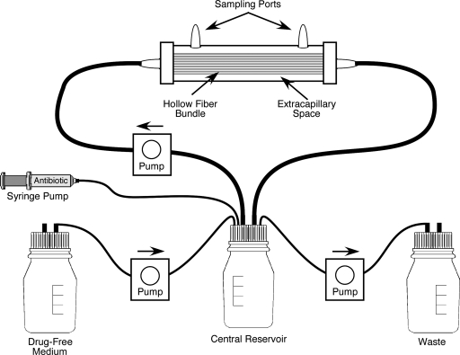
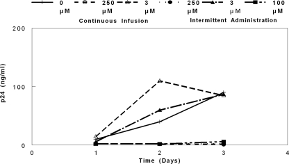
Variola major virus remains a potent weapon of bioterror. There is currently an investigational-new-drug application for cidofovir for the therapy of variola major virus infections. Stittelaar and colleagues compared the levels of effectiveness of postexposure smallpox vaccination (Elstree-RIVM) and antiviral treatment with cidofovir or an acyclic nucleoside phosphonate analogue 6-[2-(phosphonomethoxy)alkoxy]-2,4-diaminopyrimidine (HPMPO-DAPy) after lethal intratracheal infection of cynomolgus monkeys with monkeypox virus, a variola virus surrogate. Their results demonstrated that either compound was more effective than vaccination with the Ellstree vaccine (K. J. Stittelaar et al., Nature 439:745-748, 2006). An unanswered question is how to translate this information into therapy for poxvirus infections in people. In a proof-of-principle study, we used a novel in vitro hollow-fiber infection model system to determine the pharmacodynamics of vaccinia virus infection of HeLa-S3 cells treated with cidofovir. Our results demonstrate that the currently licensed dose of cidofovir of 5 mg/kg of body weight weekly with probenecid (which ameliorates nephrotoxicity) is unlikely to provide protection for patients intentionally exposed to Variola major virus. We further demonstrate that the antiviral effect is independent of the schedule of drug administration. Exposures (area under the concentration-time curve) to cidofovir that will have a robust protective effect will require doses that are 5 to 10 times that currently administered to humans. Such doses may cause nephrotoxicity, and therefore, approaches that include probenecid administration as well as schedules of administration that will help ameliorate the uptake of cidofovir into renal tubular epithelial cells need to be considered when addressing such treatment for people.


Figures

FIG. 1.
FIG. 1.
HFIM system. Each hollow-fiber cartridge contains semipermeable hollow fibers which allow gases and small-molecular-weight nutrients to pass through the membranes while keeping cells and viruses outside the membranes. Uninfected and virus-infected cells are added to the cartridge through one of the sampling ports on the top of the cartridge. Medium from the reservoir is pumped through the hollow fibers to nourish the cells that grow outside of the hollow fibers. The contents of the ECS of the hollow-fiber units are sampled for cells, cell-free virus, and drug from the ports on the top of each unit, and the concentration of drug entering the hollow-fiber unit can be determined by sampling the medium as it enters the hollow-fiber unit.
FIG. 2.
FIG. 2.
Growth of VV in the HFIM system. For growth of VV in HeLa-S3 cells, 106 VV-infected HeLa-S3 cells were mixed with 108 uninfected HeLa-S3 cells and placed in hollow-fiber units. The units were connected to reservoirs containing SMEM. The hollow-fiber units were placed in incubators at 36°C with 5% CO2, and medium was circulated through each hollow fiber for 4 days. At various times, the cells and virus produced in the ECS were sampled through the ports at the top of the cartridge. The cells were removed from the sample by centrifugation, and the amount of virus in the clarified supernatant was determined by plaque assay. The data show that all three strains of VV grow well in the HFIM system. The data are from a single experiment that is representative of several independent experiments (n = 3).
FIG. 3.
FIG. 3.
Dose-range experiment for VV and cidofovir. To determine the dose of cidofovir that will inhibit the growth of VV in the HFIM system, six hollow-fiber units were set up as described in the legend of Fig. 2 and the stated concentrations of cidofovir were continuously infused through the different units. The ECS was sampled at the indicated times, and the effect of cidofovir on virus replication was determined by plaque assay. The results show that concentrations of cidofovir equal to or greater than 50 μM inhibited VV replication in HeLa-S3 cells. The data are the result of a single experiment.
FIG. 4.
FIG. 4.
Dose fractionation experiment for VV and cidofovir. To determine the pharmacodynamically linked variable for cidofovir for VV, 108 uninfected HeLa-S3 cells and 106 HeLa-S3 cells infected with the recombinant WR strain of VV were placed into five hollow-fiber units. One unit received no cidofovir. One unit received 250 μM cidofovir as a continuous infusion. One unit received 3 μM cidofovir as a continuous infusion. One unit received 250 μM cidofovir delivered as an intermittent infusion over a 1-h period, followed by a no-drug washout. One unit received 3 μM cidofovir delivered as an intermittent infusion over 1 h, followed by a no-drug washout. One unit received 100 μM cidofovir delivered as an intermittent infusion over a 1-h period and then a no-drug washout. Each hollow-fiber unit was sampled at the indicated times, and the amount of virus produced was measured by p24 ELISA. The data show that 3 μM cidofovir delivered as an intermittent infusion or as a continuous infusion had no effect on the production of VV but that 250 μM cidofovir delivered as an intermittent infusion or as a continuous infusion inhibited virus replication, as did a bolus of 100 μM cidofovir. The data presented are the results of a single experiment.
FIG. 5.
FIG. 5.
Pharmacokinetic analysis of the dose fractionation experiment. The medium entering each hollow-fiber unit was sampled at the indicated times, and the amount of cidofovir was determined by LC-MS. The data show that the two continuous doses (3 μM and 250 μM) were maintained over the 72-h time course and that three bolus doses (3 μM, 100 μM, and 250 μM) peaked at the end of the 1-h infusion and then declined with the no-drug washout. These results demonstrate that each hollow-fiber unit actually received the intended dose of cidofovir. Contin Inf, continuous infusion.
FIG. 6.
FIG. 6.
Repeat of the dose fractionation study. Since the original dose fractionation study (Fig. 4) indicated that low doses (3 μM) given as a continuous infusion or as an intermittent administration followed by a no-drug washout had no effect on VV replication in HeLa-S3 cells, the dose fractionation study was repeated at higher doses. The results show that 30 μM drug given as a continuous infusion or as an intermittent administration followed by a no-drug washout had the same antiviral effect. Similar results occurred when 100 μM drug was given as a continuous infusion or as an intermittent administration followed by a no-drug washout. These results show that the pharmacodynamically linked variable for cidofovir is the AUC0-72/EC50 ratio. These results are from a single representative experiment.
FIG. 7.
FIG. 7.
Relationship between the antiviral effect and the measured AUC0-72. The antiviral activity as measured by p24 output from the WR strain of VV at 72 h was the dependent variable, and the AUC0-72 of cidofovir was the independent variable. The AUC0-72 required to achieve 50% of the Emax was 2,804 μM·h.

Similar articles

Cited by

References

    1. Baker, R. O., M. Bray, and J. W. Huggins. 2003. Potential antiviral therapeutics for smallpox, monkeypox and other orthopoxvirus infections. Antivir. Res. 57:13-23. - PMC - PubMed
    1. Bilello, J. A., G. Bauer, M. N. Dudley, G. A. Cole, and G. L. Drusano. 1994. Effect of 2′,3′-didehydro-3′-deoxythymidine in an in vitro hollow-fiber pharmacodynamic model system correlates with results of dose-ranging clinical studies. Antimicrob. Agent Chemother. 38:1386-1391. - PMC - PubMed
    1. Bray, M. 2003. Pathogenesis and potential antiviral therapy for complications of smallpox vaccination. Antivir. Res. 58:101-114. - PubMed
    1. Bray, M., M. Martinez, D. F. Smee, D. Kefauver, E. Thompson, and J. W. Huggins. 2000. Cidofovir protects mice against lethal aerosol or intranasal cowpox virus challenge. J. Infect. Dis. 181:10-19. - PubMed
    1. Breman, J. G., and I. Arita. 1980. The confirmation and maintenance of small pox eradication. N. Engl. J. Med. 303:1263-1273. - PubMed

Publication types