. 2008 Dec;28(24):7427-41.
doi: 10.1128/MCB.01962-07. Epub 2008 Oct 13.

Stabilized beta-catenin functions through TCF/LEF proteins and the Notch/RBP-Jkappa complex to promote proliferation and suppress differentiation of neural precursor cells

Affiliations

Stabilized beta-catenin functions through TCF/LEF proteins and the Notch/RBP-Jkappa complex to promote proliferation and suppress differentiation of neural precursor cells

Takeshi Shimizu et al. Mol Cell Biol. 2008 Dec.

Abstract

The proliferation and differentiation of neural precursor cells are mutually exclusive during brain development. Despite its importance for precursor cell self renewal, the molecular linkage between these two events has remained unclear. Fibroblast growth factor 2 (FGF2) promotes neural precursor cell proliferation and concurrently inhibits their differentiation, suggesting a cross talk between proliferation and differentiation signaling pathways downstream of the FGF receptor. We demonstrate that FGF2 signaling through phosphatidylinositol 3 kinase activation inactivates glycogen synthase kinase 3beta (GSK3beta) and leads to the accumulation of beta-catenin in a manner different from that in the Wnt canonical pathway. The nuclear accumulated beta-catenin leads to cell proliferation by activating LEF/TCF transcription factors and concurrently inhibits neuronal differentiation by potentiating the Notch1-RBP-Jkappa signaling pathway. beta-Catenin and the Notch1 intracellular domain form a molecular complex with the promoter region of the antineurogenic hes1 gene, allowing its expression. This signaling interplay is especially essential for neural stem cell maintenance, since the misexpression of dominant-active GSK3beta completely inhibits the self renewal of neurosphere-forming stem cells and prompts their neuronal differentiation. Thus, the GSK3beta/beta-catenin signaling axis regulated by FGF and Wnt signals plays a pivotal role in the maintenance of neural stem/precursor cells by linking the cell proliferation to the inhibition of differentiation.


Figures
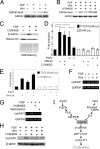
FIG. 1.
FIG. 1.
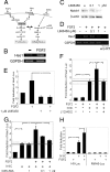
Nuclear β-catenin accumulation and induction of cyclin D1 expression through FGF2-mediated PI3K activation and GSK3β inactivation in neural precursor cells. (A) The upper panel shows the Western blot analysis with anti-phospho-GSK3β-Ser9 antibody. The lower panel shows the total amount of GSK3β examined with anti-GSK3β antibody. The upper of the two bands appeared to be phosphorylated GSK3β. Neither FGF2 nor Wnt3a affected the β-actin protein level (data not shown). (B) The PI3K inhibitor LY294002 (50 μM) impaired the GSK3β Ser9 phosphorylation induced by FGF2 treatment (20 ng/ml; n = 6). LY294002 slightly reduced GSK3β Ser9 phosphorylation induced by Wnt3a treatment (150 ng/ml; n = 3). (C) Accumulation of β-catenin was detected in the nuclear fraction of neural precursor cells treated with 20 ng/ml FGF2 for 1 h. This accumulation was blocked by 50 μM LY294002. The amounts and purities of the nuclear fraction were confirmed by Coomassie brilliant blue staining, the expression level of histone H3, and leukemia inhibitory factor receptor (LIFR; data not shown). (D) 7×TCF-SV40 promoter activity (black bars) and SV40 promoter activity (white bars) were investigated by a dual luciferase reporter assay. The height of each bar indicates the increase of luciferase activity compared to that of unstimulated cells. rmWnt-3a (100 ng/ml) was applied as a positive control for 7×TCF-Luc reporter activity (n = 3; *, P < 0.05; **, P < 0.001; Student's t test). (E) Coapplication of 20 ng/ml FGF2 and 100 ng/ml rmWnt-3a displayed an additive effect on 7×TCF promoter activation (n = 3). The height of each bar indicates the increase of luciferase activity compared to that of unstimulated cells. (F) FGF2 (20 ng/ml) and rmWnt-3a (200 ng/ml) increased the expression of cyclin D1 mRNA, as detected by RT-PCR. (G) LY294002 (50 μM) and wortmannin (100 nM) impaired FGF2-induced cyclin D1 mRNA expression (n = 3). (H) Western blot analysis with anti-cyclin D1 antibody. LY294002 (50 μM) abolished the induction of cyclin D1 by FGF2 (20 ng/ml; n = 3) but did not affect that by rmWnt-3a (150 ng/ml; n = 2). All experiments in this figure were done with cells seeded at a high density (3.2 × 10⁵ cells/cm2). (I) A schematic drawing summarizing the signaling pathways analyzed in this figure.
FIG. 2.
FIG. 2.
Involvement of PI3K activation and GSK3β Ser9 phosphorylation in the proliferation of neural precursor cells in response to FGF2. (A to D) FGF2 (10 ng/ml) increased the number of BrdU+ cells (red in panel A), and LY294002 abolished this effect in a dose-dependent manner (B). DMSO, dimethylsulfoxide. (C) Ratio of BrdU+ cells/total cells (n = 4; *, P < 0.01; **, P < 0.001; Student's t test). (D) Ratio of active caspase 3+ (casp3+) cells/total cells (n = 4). (E to G) Ki67+ proliferative cells (red in panels E and F) were less frequent among GSK3βS9A-expressing cells (green in panel F) than among control GFP virus-infected cells (green in panel E). (G) The ratio of Ki67+ cells/GFP+ cells (n = 5; *, P < 0.01 compared to values for the control; Student's t test). All of these experiments were done with cells seeded at a high density (3.2 × 10⁵ cells/cm2). (H to L) Misexpression of GSK3βS9A significantly decreased the diameter of primary neurospheres (green sphere in panel K) compared to that of the control (green sphere in panel J; also see the corresponding uncolored spheres in panels H and I). Yellow dots seen in panel K are artifacts caused by the reflection of the lights from clean benches. (L) Summary of results (n = 4). Scale bar for panels A and B, 100 μm. Scale bar for panels E and F, 50 μm. Scale bar for panels H to K, 100 μm.
FIG. 3.
FIG. 3.
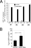
Defect in secondary sphere formation by dominant-active GSK3β-expressing cells. (A) GSK3βS9A-GFP- or GFP-retrovirus-infected cells in primary neurospheres were dissociated and used for a secondary sphere assay. GSK3βS9A-expressing cells (S9A) formed no or very few secondary spheres compared to control GFP-expressing cells (cont). The numbers at the bottom indicate each of the four independent experiments. (B) Neural precursor cells were infected with GSK3βS9A-GFP or GFP virus, and primary neurospheres formed after 8 days were dissociated and replated on 8-well chamber slides. Six hours later, the number of Tuj1+ cells among GSK3βS9A-GFP-expressing cells and that among GFP-expressing cells were counted (n = 3; *, P < 0.05 compared to control values; Student's t test).
FIG. 4.
FIG. 4.
Potentiation of Notch-mediated hes promoter activation by FGF2 signaling via PI3K activation and GSK3β inactivation. (A) Our working hypothesis. FGF signaling and Notch signaling cooperate to regulate cell fate decisions. (B) FGF2 treatment (20 ng/ml) of neural precursor cells for 3 h increased the level of Hes1 mRNA, as detected by RT-PCR. PCR products were not amplified from samples without reverse transcriptase treatment (data not shown). (C) γ-Secretase inhibitor L685458 decreased the amount of N1IC in a high density of neural precursor cells cultured for 3 days. (D) L685458 abolished FGF2-induced hes1 gene expression in a high-cell-density culture of neural precursor cells examined by RT-PCR. The hes1 mRNA level was increased by a 3-h FGF2 treatment; however, the FGF2 effect was abolished by the addition of 1 μM L685458. No band was detected without reverse transcriptase (w/o RT). (E) L685458 (1 μM) abolished FGF2-induced hes1 gene promoter activation. Neural precursor cells cultured at a high cell density were transfected with a plasmid containing pHes1-Luc. hes1 promoter activity was investigated by a dual luciferase reporter assay. Each bar indicates the increase of luciferase activity compared to that of untreated cells (n = 3; *, P < 0.001; Student's t test). (F) Neural precursor cells cultured at a low cell density were cotransfected with plasmid containing pHes1-Luc with or without the cDNA encoding N1IC. FGF2 (20 ng/ml) potentiated hes1 promoter activity only when the cells expressed N1IC. LY294002 treatment (50 μM) abolished FGF2-induced potentiation of hes1 promoter activity (n = 5; *, P < 0.05; **, P < 0.01; ***, P < 0.001; Student's t test). (G) Misexpression of GSK3βS9A blocked the ability of FGF2 to potentiate N1IC-induced hes1 promoter activation (n = 4; *, P < 0.05; **, P < 0.01; Student's t test). (H) Neural precursor cells were transfected with N1IC cDNA together with H5-Luc or RMH5-Luc with mutations in the RBP-Jκ binding site. (n = 3; ***, P < 0.001; Student's t test).
FIG. 5.
FIG. 5.
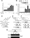
Novel function of β-catenin as a transcriptional coactivator for Notch signaling. (A) Expression of stabilized (stb) β-catenin potentiated N1IC-induced hes1 promoter activation in neural precursor cells in a dose-dependent manner. The misexpression of dominant-negative human TCF4 (ΔN-hTCF4) did not affect hes1 promoter activity (n = 4; *, P < 0.01; **, P < 0.001; Student's t test). (B) The ectopic expression of HA-tagged β-catenin (βcat) was potently inhibited by the cotransfection of NIH 3T3 cells with β-catenin siRNA (siR). (C) β-Catenin siRNA abolished the effect of FGF2 on hes1 promoter activation, whereas it did not affect N1IC-mediated hes1 promoter activation (n = 4; *, P < 0.01; **, P < 0.001; Student's t test). (D) Endogenous β-catenin and Notch1 proteins in neural precursor cells were coimmunoprecipitated. Preincubation of anti-Notch1 antibody (αNotch1) with its blocking peptide (B.P.) prevented the coprecipitation of β-catenin and N1IC. The β-catenin signal density was quantified with densitometry, and the result is indicated under each band. IP, immunoprecipitation. (E and F) The molecular interaction between N1IC and β-catenin was analyzed by coimmunoprecipitation assays in HEK293 cells that overexpressed the cDNAs for HA-tagged β-catenin and myc-tagged N1IC. Anti-HA (αHA) antibody was used for immunoprecipitation, and anti-myc antibody was used for the Western blotting analysis shown in panel E. Anti-myc antibody (αmyc) was used for immunoprecipitation, and anti-HA antibody was used for the Western blotting analysis shown in panel F. (G) ChIP assay demonstrated that β-catenin is associated with the hes1 promoter region in neural precursor cells. Chromosomal DNA was precipitated with normal rabbit IgG from Upstate (lane 1), normal rabbit IgG from Santa Cruz Biotechnology (lane 2), or anti-β-catenin antibody (α β-catenin) (lanes 3, 4, and 5). Chromosomal DNA was prepared from high-density neural precursor cells (lanes 1, 2, 3, and 4), from the cells treated with 1 μM L685458 (lane 4), or from low-density cultures of neural precursor cells (lane 5). Arrows in the drawing indicate PCR primer positions.
FIG. 6.
FIG. 6.
Physical and functional interactions among N1IC, β-catenin, p300, and P/CAF in hes1 promoter activation. (A and B) HEK293 cells were transfected with expression vectors containing tagged cDNAs. The molecular interaction between N1IC and P/CAF was facilitated in the presence of both p300 and β-catenin (βcat). Furthermore, β-catenin was efficiently immunoprecipitated with N1IC in the presence of both p300 and P/CAF (B). IP; αCont IgG, immunoprecipitation with anti-control IgG. (C) The contribution of endogenous p300 to hes1 promoter activation in neural precursor cells was examined using truncated forms of dominant-negative p300 (n = 3; *, P < 0.05; **, P < 0.01; ***, P < 0.001; Student's t test). stb, stabilized. (D) Neural precursor cells were transfected with combinations of pHes1-Luc, N1IC, stabilized β-catenin, and P/CAF and p300 cDNAs (n = 4; *, P < 0.05; **, P < 0.01; Student's t test). αmyc, anti-myc antibody.
FIG. 7.
FIG. 7.
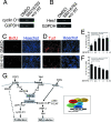
Correlation of GSK3β activity with cell proliferation and neuronal differentiation. (A and B) SB216763 (5 μM) treatment of neural precursor cells for 3 h increased the amount of cyclin D1 that had not been amplified from samples without reverse transcriptase (w/o RT) in the reaction mixture. (C to F) SB216763 treatment of neural precursor cells increased the number of BrdU+ cells (red in panel C) and decreased the number of Tuj1+ cells (red in panel D). The ratios of BrdU+ cells/total cells and Tuj1+ cells/total cells are shown in panels E and F, respectively (n = 3; *, P < 0.05, **; P < 0.01; Student's t test). (G) Schematic drawing summarizing our model. β-Catenin plays an important role in linking the promotion of proliferation and the inhibition of differentiation in neural precursor cells by coordinating signals initiated by FGF2, canonical Wnts, and Notch ligands. The inset represents the protein complex governing hes1 promoter activation. BS, binding site; DMSO, dimethylsulfoxide.

Similar articles

Cited by

References

    1. Alves-Guerra, M. C., C. Ronchini, and A. J. Capobianco. 2007. Mastermind-like 1 is a specific coactivator of beta-catenin transcription activation and is essential for colon carcinoma cell survival. Cancer Res. 678690-8698. - PubMed
    1. Amit, M., M. K. Carpenter, M. S. Inokuma, C. P. Chiu, C. P. Harris, M. A. Waknitz, J. Itskovitz-Eldor, and J. A. Thomson. 2000. Clonally derived human embryonic stem cell lines maintain pluripotency and proliferative potential for prolonged periods of culture. Dev. Biol. 227271-278. - PubMed
    1. Andjelkoviæ, M., D. R. Alessi, R. Meier, A. Fernandez, N. J. Lamb, M. Frech, P. Cron, P. Cohen, J. M. Lucocq, and B. A. Hemmings. 1997. Role of translocation in the activation and function of protein kinase B. J. Biol. Chem. 27231515-31524. - PubMed
    1. Arcaro, A., and M. P. Wymann. 1993. Wortmannin is a potent phosphatidylinositol 3-kinase inhibitor: the role of phosphatidylinositol 3,4,5-trisphosphate in neutrophil responses. Biochem. J. 296297-301. - PMC - PubMed
    1. Belham, C., S. Wu, and J. Avruch. 1999. Intracellular signalling: PDK1-a kinase at the hub of things. Curr. Biol. 9R93-R96. - PubMed

Publication types

MeSH terms