Comparative Study
. 2008 Oct 27;205(11):2483-90.
doi: 10.1084/jem.20080039. Epub 2008 Oct 13.

Stromal mesenteric lymph node cells are essential for the generation of gut-homing T cells in vivo

Affiliations
Comparative Study

Stromal mesenteric lymph node cells are essential for the generation of gut-homing T cells in vivo

Swantje I Hammerschmidt et al. J Exp Med. .

Abstract

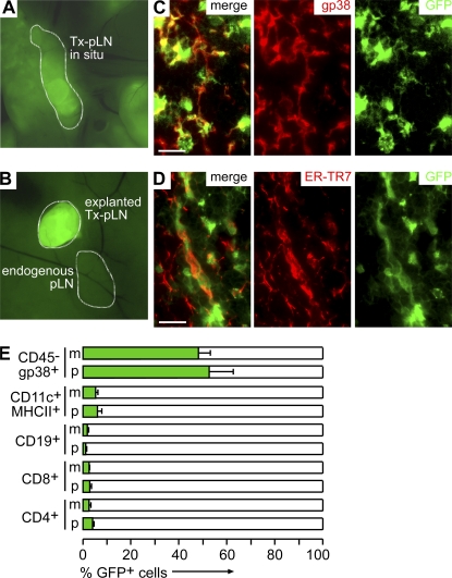
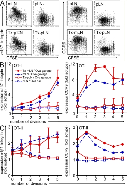
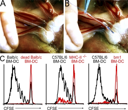
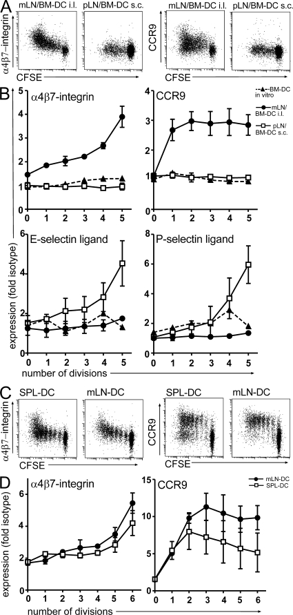
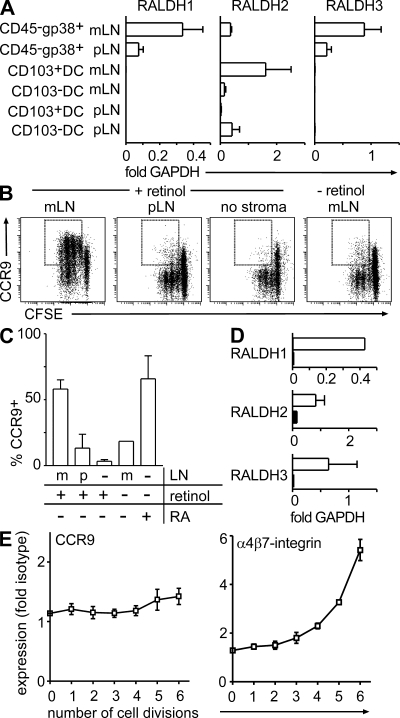
T cells primed in the gut-draining mesenteric lymph nodes (mLN) are imprinted to express alpha4beta7-integrin and chemokine receptor CCR9, thereby enabling lymphocytes to migrate to the small intestine. In vitro activation by intestinal dendritic cells (DC) or addition of retinoic acid (RA) is sufficient to instruct expression of these gut-homing molecules. We report that in vivo stroma cells, but not DC, allow the mLN to induce the generation of gut tropism. Peripheral LN (pLN) transplanted into the gut mesenteries fail to support the generation of gut-homing T cells, even though gut-derived DC enter the transplants and prime T cells. DC that fail to induce alpha4beta7-integrin and CCR9 in vitro readily induce these factors in vivo upon injection into mLN afferent lymphatics. Moreover, uniquely mesenteric but not pLN stroma cells express high levels of RA-producing enzymes and support induction of CCR9 on activated T cells in vitro. These results demonstrate a hitherto unrecognized contribution of stromal cell delivered signals, including RA, on the imprinting of tissue tropism in vivo.


Figures

Figure 1.
Figure 1.
Transplantation of LN fragments yields chimeric LN constituted by donor-derived stromal cells and recipient-derived hematopoietic cells. (A) 8 wk after transplantation, Tx-pLN and Tx-mLN could be identified in situ by expression of EGFP. (B) To demonstrate low autofluorescence in endogenous LN, an excised Tx-pLN was placed besides the endogenous pLN. (C and D) Tx-pLN were analyzed by fluorescence microscopy for EGFP expression (green). Expression of gp38 (C) and ER-TR7 (D) is depicted in red. Comparable results were obtained for Tx-mLN (not depicted). Bars, 20 μm. (E) Cells were isolated from Tx-LN, and EGFP expression by stroma cells, DC, B, and T cells was analyzed by flow cytometry (data are pooled from four mice analyzed in two experiments). Error bars represent SD.
Figure 2.
Figure 2.
Heterotopic chimeric Tx-pLN, but not orthotopic Tx-mLN, fail to generate gut-homing T cells in vivo. Cells isolated from OT-I or OT-II Ly5.1 mice were labeled with CFSE and adoptively transferred into mice that received Tx-pLN or Tx-mLN 8 wk before. 1 d later, a single dose of Ova was applied orally or injected s.c. (A) Representative results obtained for α4β7-integrin and CCR9 expression by OT-I T cells (DAPIVα2+Vβ5+CD8+) activated in the mLN, Tx-mLN, and Tx-pLN after oral antigen application and in pLN after s.c. injection of antigen. (B and C) Diagrams depict expression of α4β7-integrin and CCR9 as fold isotype control for OT-I T cells (DAPIVα2+Vβ5+CD8+; B) and OT-II T cells (DAPILy5.1+Vβ5+CD4+; C). All experiments have been performed at least three times with two or more mice per group. Error bars represent SD.
Figure 3.
Figure 3.
Injection of antigen-loaded BM-DC into mLN afferent lymphatics induces proliferation of antigen-specific T cells. Recipient mice received CFSE-labeled LN cells isolated from DO11.10, OT-I, or OT-II donors. 1 d later, these mice received oil by gavage 1 h before surgery to visualize mLN afferent lymphatics and to facilitate i.l. injection. (A) The small intestine was exposed by surgery and mLN afferent lymphatics were identified by their white color. (B) To demonstrate the method of i.l. injection, ∼0.3 μl of blue dye was injected into a single lymph vessel. After injection of 2× 1 μl into two separate vessels, as performed for routine injection of DC, the fluid spread throughout the LN sinus (not depicted). (C) 105 BM-DC was injected into two separate lymphatic vessels opening into the distal aspect of the mLN chain. Injection of Ova loaded DC (solid black line), but not heat-killed DC (red line), induced proliferation of Ova-specific DO11.10 T cells (DAPIKJ16-26+CD4+). BM-DC derived from MHC class II–deficient mice induced only marginal proliferation of OT-II cells. Similarly, DC derived from BM of bm-1 mice failed to activate OT-I T cells. All experiments have been performed at least two times with three or more mice per group.
Figure 4.
Figure 4.
BM-DC and spleen-derived DC generate gut-homing T cells in vivo but not in vitro. (A and B) DO11.10 cells were adoptively transferred into WT recipients. 1 d later, BM-DC were either injected i.l. or s.c., and proliferating DO11.10 T cells in the mLN and pLN were analyzed for expression of α4β7-integrin, CCR9, and E- and P-selectin ligand as indicated. Moreover, expression of these molecules was assessed after in vitro coculture of antigen-loaded BM-DC with DO11.10 T cells at a 1:10 ratio. BM-DC injected into mLN afferent lymphatics, but not injected s.c., or in vitro cocultures led to the up-regulation of α4β7-integrin and CCR9 on proliferating T cells. Conversely, s.c., but not i.l., injection of BM-DC induced expression of E- and P-selectin ligand. (C and D) DC were isolated from mLN and spleen, loaded in vitro with Ova peptide, and injected i.l. into recipient mice as described in Fig. 3. Spleen-derived DC that fail to up-regulate gut-homing factors after 3 d of coculture with DO11.10 cells in vitro (not depicted) readily induced expression of α4β7-integrin and modest expression of CCR9 after i.l. injection in vivo. All experiments were performed at least three times with at least two mice per group.
Figure 5.
Figure 5.
mLN- but not pLN-derived stroma cells express high levels of RALDH and support the induction of CCR9 on proliferating T cells. (A) cDNA was prepared from sorted CD45CD24gp38+ stroma cells and CD103+ as well as CD103 DC (CD11c+MHCII+) purified from pLN and mLN. Expression of RALDH1, 2, and 3 was assessed by real-time PCR and is depicted as fold expression compared with GAPDH. Data depict the mean and SD of three to five independent experiments measured in duplicates. (B and C) CFSE-labeled OT-I cells were activated by anti-CD3/CD28 treatment in the presence of either pLN- or mLN-derived stroma cells with or without addition of retinol. Stroma cells were enriched by adherence and culture of digested LN cell suspensions over 10 d. Data shown represent the mean and SD of three independent experiments. (D) cDNA was prepared from sorted CD45CD24gp38+ stroma cells (open bars) and CD103+ DC (closed bars) of CCR7-deficient mLN. Expression of RALDH1, 2, and 3 was assessed by real-time PCR. Bars depict the mean and SD of fold expression compared with GAPDH observed in two independent experiments. (E) CCR9 expression was analyzed after i.l. injection of antigen-loaded BM-DC into mLN afferent lymphatics of CCR7-deficient mice. In contrast to the situation in WT mice, BM-DC failed to support the induction of CCR9 on T cells in CCR7-deficient mice. Expression of α4β7-integrin was not significantly affected. Results depict data obtained in one out of two experiments performed with five mice. Error bars represent SD.

References

    1. McDermott, M.R., and J. Bienenstock. 1979. Evidence for a common mucosal immunologic system. I. Migration of B immunoblasts into intestinal, respiratory, and genital tissues. J. Immunol. 122:1892–1898. - PubMed
    1. Cahill, R.N., D.C. Poskitt, D.C. Frost, and Z. Trnka. 1977. Two distinct pools of recirculating T lymphocytes: migratory characteristics of nodal and intestinal T lymphocytes. J. Exp. Med. 145:420–428. - PMC - PubMed
    1. Agace, W.W. 2006. Tissue-tropic effector T cells: generation and targeting opportunities. Nat. Rev. Immunol. 6:682–692. - PubMed
    1. Wagner, N., J. Lohler, E.J. Kunkel, K. Ley, E. Leung, G. Krissansen, K. Rajewsky, and W. Müller. 1996. Critical role for beta7 integrins in formation of the gut-associated lymphoid tissue. Nature. 382:366–370. - PubMed
    1. Lefrancois, L., C.M. Parker, S. Olson, W. Muller, N. Wagner, M.P. Schon, and L. Puddington. 1999. The role of β7 integrins in CD8 T cell trafficking during an antiviral immune response. J. Exp. Med. 189:1631–1638. - PMC - PubMed

Publication types

MeSH terms