Comparative Study
. 2008 Oct 27;205(11):2643-55.
doi: 10.1084/jem.20080730. Epub 2008 Oct 13.

CXCL12 (SDF-1alpha) suppresses ongoing experimental autoimmune encephalomyelitis by selecting antigen-specific regulatory T cells

Affiliations
Comparative Study

CXCL12 (SDF-1alpha) suppresses ongoing experimental autoimmune encephalomyelitis by selecting antigen-specific regulatory T cells

Moran Meiron et al. J Exp Med. .

Abstract

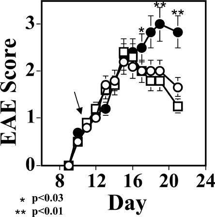
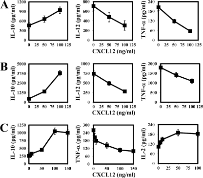
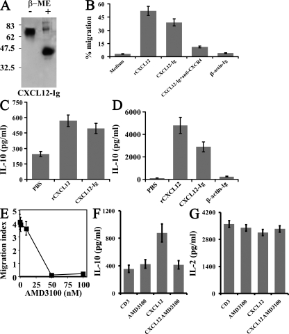
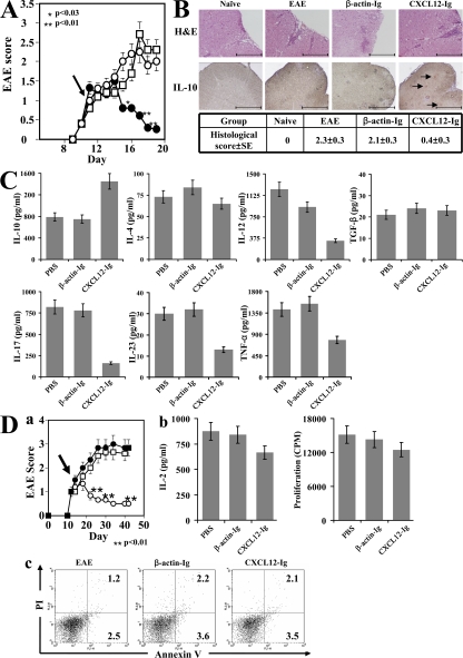
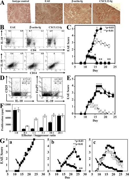
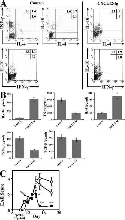
Experimental autoimmune encephalomyelitis (EAE) is a T cell-mediated autoimmune disease of the central nervous system induced by antigen-specific effector Th17 and Th1 cells. We show that a key chemokine, CXCL12 (stromal cell-derived factor 1alpha), redirects the polarization of effector Th1 cells into CD4(+)CD25(-)Foxp3(-)interleukin (IL) 10(high) antigen-specific regulatory T cells in a CXCR4-dependent manner, and by doing so acts as a regulatory mediator restraining the autoimmune inflammatory process. In an attempt to explore the therapeutic implication of these findings, we have generated a CXCL12-immunoglobulin (Ig) fusion protein that, when administered during ongoing EAE, rapidly suppresses the disease in wild-type but not IL-10-deficient mice. Anti-IL-10 neutralizing antibodies could reverse this suppression. The beneficial effect included selection of antigen-specific T cells that were CD4(+)CD25(-)Foxp3(-)IL-10(high), which could adoptively transfer disease resistance, and suppression of Th17 selection. However, in vitro functional analysis of these cells suggested that, even though CXCL12-Ig-induced tolerance is IL-10 dependent, IL-10-independent mechanisms may also contribute to their regulatory function. Collectively, our results not only demonstrate, for the first time, that a chemokine functions as a regulatory mediator, but also suggest a novel way for treating multiple sclerosis and possibly other inflammatory autoimmune diseases.


Figures

Figure 1.
Figure 1.
Neutralization of CXCL12 during ongoing EAE aggravates ongoing EAE. C57BL/6 female mice (n = 6 per group) were subjected to active induction of MOGp35-55-induced EAE, and at the onset of disease (day 10) were separated into three equally sick groups (n = 6 mice per group). On days 11, 13, 15, and 17 after the induction of disease, mice were injected i.v. either with PBS (open circles), 50 μg/mouse of anti-CXCL12 mAb (closed circles), or control antibody (open squares). An observer blind to the experimental protocol monitored the development and progression of disease. The results of one out of three independent experiments (n = 6 mice per each group) are shown as the mean maximal score ± SE. The arrow indicates the first day of anti-CXCL12 antibody administration.
Figure 2.
Figure 2.
CXCL12 directs the functional polarization of macrophages and T cells into high IL-10, low inflammatory mediator–producing cells. (A–C) CXCL12 was added at different concentrations to primary whole spleen culture taken from EAE mice and stimulated with their target MOGp35-55 antigen for 72 h (A), freshly isolated peritoneal macrophages stimulated with 0.5 μg/ml LPS for 48 h (B), or purified naive CD4+ T cells activated with anti-CD3/anti-CD28 for 48 h (C). Cytokine concentrations were measured in triplicates using a standard ELISA method. Results shown in this figure represent three independent experiments with similar results and are presented as means ± SE.
Figure 3.
Figure 3.
CXCL12-Ig preserves the biological activities of native CXCL12. (A) Purified CXCL12-Ig was separated on 12% SDS-PAGE and subjected to Western blot analysis under reducing and nonreducing conditions (with or without β-mercaptoethanol) using anti-CXCL12 mAb (clone 79014) as a primary antibody (molecular masses are shown). (B) THP-1 cells (human monocytic cell line) were subjected to a migration assay using a Transwell system. Lower chambers were supplemented with culture medium, rCXCL12, CXCL12-Ig, CXCL12-Ig plus anti-CXCR4 mAb, or β-actin–Ig. The number of cells migrating to the lower chamber was counted by FACS 3 h later. Results shown represent three independent experiments and are the mean of the migration percentage (number of cells that migrated to the lower chamber divided by the number of cells originally plated in the upper chamber) ± SE. (C) Freshly isolated peritoneal macrophages were supplemented with PBS, rCXCL12, or CXCL12-Ig. Supernatants were collected 48 h later, and the IL-10 concentration was determined by ELISA. The results shown represent three experiments done in triplicates, and are the mean IL-10 concentration ± SE. (D) Primary spleen cell cultures responding to their target MOGp35-55 antigen were supplemented with PBS, rCXCL12, CXCL12-Ig, or β-actin–Ig. Supernatants were collected 48 h later, and IL-10 levels were determined by standard ELISA. The results represent three experiments done in triplicates, and are the mean IL-10 concentration ± SE. (E) Dose-dependent inhibition of CXCL12-induced migration of anti-CD3/anti-CD28–activated spleen T cells from naive C57BL/6 mice. Results are shown as the mean ± SE of three independent experiments with similar results. (F and G) IL-10 and IL-2 production of anti-CD3/anti-CD28–activated spleen T cells in the presence of 100 ng/ml CXCL12, 100 nM AMD3100, or CXCL12 plus 100 nM AMD3100. The results represent three experiments done in triplicates and are shown as the mean cytokine concentration ± SE.
Figure 4.
Figure 4.
CXCL12-Ig suppresses ongoing EAE. (A) C57BL/6 female mice were subjected to active induction of EAE (MOGp35-55/CFA), and just after the onset of disease (day 11), they were separated into equally sick groups (n = 6 mice each). On days 11, 13, 15, and 17, these groups were injected i.v. with either PBS (open circles), CXCL12-Ig (closed circles), or β-actin–Ig (open squares) and were monitored for the progression of disease by an observer blind to the experimental protocol. Results of one out of three independent experiments (n = 6 mice per each group) are shown as the mean maximal score ± SE. The arrow indicates the first day of CXCL12-Ig administration. (B) On day 20, three representative mice from each group were subjected to histological analysis of the lumbar spinal cord (eight sections per sample). A scale ranging from 0 to 3, based on the number of perivascular lesions per section, was used to quantify the histological score of disease, as described in Materials and methods. The table presents the quantification analysis of these sections, and a representative section from each group is also shown. Representative sections were also subjected to immunohistochemistry for IL-10. Arrows indicate cells stained positive for IL-10. Bars, 200 μm. (C) In a subsequent experiment, conducted under the same experimental protocol, mice were killed on day 15, and spleen cells from each group were cultured in the presence of their target antigen (MOGp35-55). After 24 h, levels of IL-10, IL-4, TGF-β, IL-12, IL-17, IL-23, and TNF-α were recorded by ELISA. Results are shown as the mean of triplicates ± SE. (D, a) C57BL/6 female mice were subjected to active induction of a long-term form of disease (reference 21), and just after the onset of disease they were separated into equally sick groups (n = 6 mice each). Twice a week, these groups were injected i.v. with PBS (closed circles), CXCL12-Ig (open circles), or β-actin–Ig (open squares) and monitored for the development and progression of disease by an observer blind to the experimental protocol. Results of one out of three independent experiments (n = 6 mice per each group) are shown as the mean maximal score ± SE. The arrow indicates the first day of CXCL12-Ig administration. (b) Just before the peak of disease (day 24), primary T cells from the cervical lymph nodes of PBS-, β-actin–Ig–, and CXCL12-Ig–treated mice were subjected to MOGp35-55-induced activation. The proliferative response and levels of IL-2 were recorded. (c) Apoptosis in CD4+ T cells in these cultures was determined by flow cytometry using Annexin V/PI staining (percentages are shown).
Figure 5.
Figure 5.
Antigen-specific T cells selected in the presence of CXCL12 suppress EAE. C57BL/6 female mice were subjected to active induction of EAE (MOGp35-55/CFA), and just after the onset of disease (day 11), they were separated into equally sick groups (n = 6 mice each). On days 11 and 13, these groups were injected i.v. either with PBS, CXCL12-Ig, or β-actin–Ig. On day 15, the spleens were removed. (A) Spleen sections were subjected to immunohistochemical analysis for IL-10 expression. Bars, 200 μm. (B) Spleen cells from the different groups were cultured with the target antigen for 72 h and were then subjected to flow cytometry analysis for intracellular staining of IL-10 in macrophages/dendritic cells (CD14+) and in CD4+ T cells (percentages are shown). (C) Spleen cells isolated from treated mice (in B) were subjected to antigen-specific activation and were injected (20 × 106 cells per mouse) into recipient EAE mice at the onset of disease (n = 6 mice per group) either with cells isolated from CXCL12-Ig mice (closed squares) or from β-actin–Ig–treated EAE mice (closed circles). A third group of recipients was administered with PBS (open squares). All groups were monitored for the development and progression of disease by an observer blind to the experimental protocol. Results of one out of three independent experiments (n = 6 mice per each group) are shown as the mean maximal score ± SE. (D) Before being administered to EAE mice (in C), IL-10high T cells selected in CXCL12-Ig–treated mice were tested for the expression of CD25 and FOXp3 (percentages are shown). (E) Spleen cells from EAE mice that were treated with CXCL12-Ig, as described in C, were subjected to antigen-specific in vitro activation and separated into CD4+ and CD14+ (MACS beads). 10 × 106 cells per mouse were injected into recipient EAE mice at the onset of disease (n = 6 mice per group) as follows: CD4+ cells isolated from CXCL12-Ig mice (open circles) and CD14+ cells isolated from CXCL12-Ig mice (closed circles). Control EAE mice were administered with PBS (closed squares). All groups were monitored for the development and progression of disease by an observer blind to the experimental protocol. Results of one out of three independent experiments (n = 6 mice per each group) are shown as the mean maximal score ± SE. (F) IL-10high T cells selected in CXCL12-Ig treated mice were tested for their ability to suppress the proliferative response of antigen-specific effector T cells from control EAE mice when added at different effector/regulatory ratios (shaded bars) and in the presence of 50 μg/ml of neutralizing anti–IL-10 antibody (open bars). (G) IL-10−/− mice (a) and C57BL/6 mice (b) were subjected to the treatment protocol described in Fig. 4 A. On day 13, mice were injected with either PBS (open squares), β-actin–Ig (closed circles), or CXCL12-Ig (closed squares). (c) C57BL/6 female mice were subjected to active induction of EAE, and just after the onset of disease (day 9), they were separated into equally sick groups (n = 6 mice each). On days 10, 12, and 14, mice were injected i.v. with PBS (open squares), CXCL12-Ig (closed circles), anti–IL-10 mAb (closed squares), or CXCL12-Ig followed by anti–IL-10 mAb injected 6 h later (open triangles). Mice were monitored daily for the progression of the disease by an observer blind to the treatment protocol. The arrow indicates the first day of CXCL12-Ig administration. Results of one out of three independent experiments with similar results (n = 6 mice per each group) are shown as the mean maximal score ± SE.
Figure 6.
Figure 6.
CXCL12-Ig redirects the polarization of antigen-specific effector (Th1) cells into IL-10–producing regulatory T cells that suppress EAE. (A) The MOGp35-55 CD4+ T cell line was selected during two subsequent stimulation cycles in the presence of the target antigen and the combination of recombinant mouse IL-12 and anti–IL-4–neutralizing antibodies, and were activated in the third cycle in the presence or absence of 50 μg/ml CXCL12-Ig. Cells were subjected to intracellular cytokine staining (percentages are shown). (B) Cytokine levels in the culture media were also recorded using a standard ELISA method. (C) 3 × 10⁶ T cells per mouse from the CXCL12-Ig–supplemented MOGp35-55 line (closed squares), the MOGp35-55 line (open squares), or PBS (open circles, no cells) were administered to EAE mice on day 12. Mice were monitored daily for the progression of the disease. Results of one out of two independent experiments with similar data (n = 6 mice per each group) are shown as the mean EAE score ± SE. The arrow indicates the day of cell therapy.

References

    1. Rossi, D., and A. Zlotnik. 2000. The biology of chemokines and their receptors. Annu. Rev. Immunol. 18:217–242. - PubMed
    1. Charo, I.F., and R.M. Ransohoff. 2006. The many roles of chemokines and chemokine receptors in inflammation. N. Engl. J. Med. 354:610–621. - PubMed
    1. Huang, D., Y. Han, M.R. Rani, A. Glabinski, C. Trebst, T. Sorensen, M. Tani, J. Wang, P. Chien, S. O'Bryan, et al. 2000. Chemokines and chemokine receptors in inflammation of the nervous system: manifold roles and exquisite regulation. Immunol. Rev. 177:52–67. - PubMed
    1. Karpus, W.J., N.W. Lukacs, B.L. McRae, R.M. Strieter, S.L. Kunkel, and S.D. Miller. 1995. An important role for the chemokine macrophage inflammatory protein-1 alpha in the pathogenesis of the T cell-mediated autoimmune disease, experimental autoimmune encephalomyelitis. J. Immunol. 155:5003–5010. - PubMed
    1. Homey, B., A. Muller, and A. Zlotnik. 2002. Chemokines: agents for the immunotherapy of cancer? Nat. Rev. Immunol. 2:175–184. - PubMed

Publication types

MeSH terms