. 2008 Oct 20;183(2):313-22.
doi: 10.1083/jcb.200802025. Epub 2008 Oct 13.

ODA16 aids axonemal outer row dynein assembly through an interaction with the intraflagellar transport machinery

Affiliations

ODA16 aids axonemal outer row dynein assembly through an interaction with the intraflagellar transport machinery

Noveera T Ahmed et al. J Cell Biol. .

Abstract

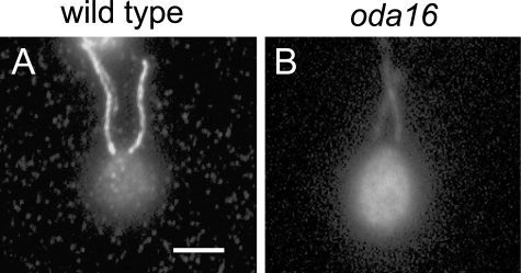
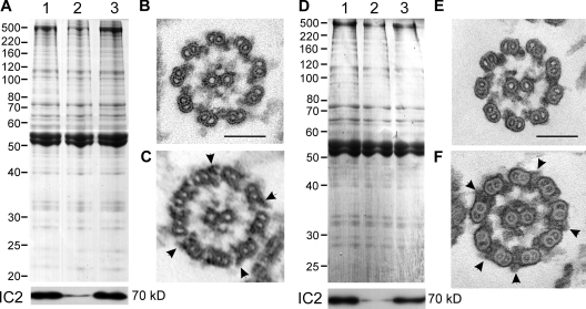
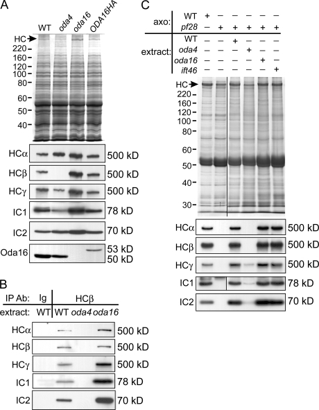
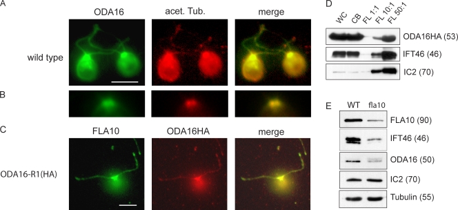
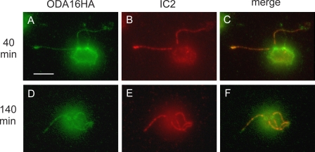
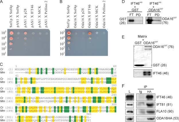
Formation of flagellar outer dynein arms in Chlamydomonas reinhardtii requires the ODA16 protein at a previously uncharacterized assembly step. Here, we show that dynein extracted from wild-type axonemes can rebind to oda16 axonemes in vitro, and dynein in oda16 cytoplasmic extracts can bind to docking sites on pf28 (oda) axonemes, which is consistent with a role for ODA16 in dynein transport, rather than subunit preassembly or binding site formation. ODA16 localization resembles that seen for intraflagellar transport (IFT) proteins, and flagellar abundance of ODA16 depends on IFT. Yeast two-hybrid analysis with mammalian homologues identified an IFT complex B subunit, IFT46, as a directly interacting partner of ODA16. Interaction between Chlamydomonas ODA16 and IFT46 was confirmed through in vitro pull-down assays and coimmunoprecipitation from flagellar extracts. ODA16 appears to function as a cargo-specific adaptor between IFT particles and outer row dynein needed for efficient dynein transport into the flagellar compartment.


Figures

Figure 1.
Figure 1.
Outer arm dynein distribution in the absence of ODA16. Immunofluorescence images of wild-type (A) and oda16 (B) cells stained with anti–ODA-IC2 show uniform distribution of outer arm dynein along the length of the flagella in both strains, although signal intensity is lower in the mutant flagella. Cytoplasmic distribution of ODA-IC2 is also similar in both strains, but abundance appears greater in mutant cytoplasm. Bar, 5 μm.
Figure 2.
Figure 2.
Outer arm dynein binds onto oda16 axonemes in vitro. (A) A Coomassie blue–stained SDS-PAGE gel of wild-type axonemes (1), oda16 axonemes (2), and oda16 axonemes combined 1:1 with a dynein-containing extract from wild type axonemes (3), and a Western blot (bottom) of the same protein samples probed with an antibody against ODA-IC2, showing restoration of outer arm dynein on the oda16 axonemes to wild-type levels. A comparison between electron micrographs of cross sections through an oda16 axoneme (B) and an oda16 axoneme incubated with a wild-type dynein extract (C) shows that a full complement of outer arm dyneins (C, arrows) were restored to the oda16 axonemes incubated with the wild-type dynein extract. The same preparations were used for the gel samples in lanes 2 and 3 of A and for the electron microscopy samples in B and C. (D) A Coomassie blue–stained SDS-PAGE gel of wild-type axonemes (lane 1), oda16 axonemes (lane 2), and oda16 axonemes treated with a dynein-containing extract from oda16 axonemes that had been concentrated threefold (lane 3), and a corresponding Western blot (bottom) was probed with an anti–ODA-IC2 antibody. Outer arm dynein has been restored on the oda16 axonemes to near-wild-type levels. Electron microscopy cross sections of the material used for lane 2 (E) and lane 3 (F) show that a nearly wild-type complement of outer arm dyneins (F, arrows) were restored on the oda16 axoneme after incubation with concentrated oda16 dynein extract. Numbers adjacent to gel blot panels indicate the estimated mass in kD of each detected band. Bars, 100 nm.
Figure 3.
Figure 3.
ODA16 is not needed for dynein preassembly in the cytoplasm. (A) Western blots demonstrate subunit abundance in wild-type and mutant cytoplasmic extracts. HCβ mutant oda4 lacks HCβ but retains normal levels of other dynein subunits and ODA16 protein. The oda16 extract lacks ODA16 and has abnormally high levels of dynein subunits. Expression of ODA16HA in the oda16 cells (ODA16HA) restores dynein subunits to wild-type levels. Note the increased size of ODA16 due to the HA tag. (B) Axonemal outer row dynein subunits are preassembled in oda16 cytoplasm. Immunoprecipitation of dynein with anti-HCβ coprecipitates all three heavy chains and both intermediate chains from both wild-type (WT) and oda16 cytoplasmic extracts. Mock precipitation from the wild type with anti-HA (Ig) and from oda4 with anti-HCβ failed to precipitate any subunits. (C) Axonemal dynein in oda16 cytoplasm is competent to bind to axonemes. Stained gels and blots of axonemes from the wild type (WT) or pf28 cells separated by SDS-PAGE show that pf28 axonemes completely lack outer row dynein (first two lanes). To test the ability of axonemal dynein in the cytoplasmic pool to bind to axonemes, pf28 axonemes and cytoplasmic extracts were incubated together at a 1:2 stoichiometric ratio for 1 h, and washed axonemes were analyzed (last four lanes). Outer arm dynein proteins in wild-type, oda16, and ift46 extracts, but not oda4 extracts, were competent to bind to pf28 axonemes and restore dynein to wild-type levels. The stained gel and IC1 blot in C used identical sample loads; the first two lanes were originally run on the same gel as other lanes, but intervening lanes have been spliced out (indicated by black vertical lines). Gels used to prepare blots of all three HCs and IC2 in C were prepared with fivefold less protein than is shown in the gel. In addition to the mass of size standards, the position of dynein heavy chains (HC) is indicated by an arrow next to the stained gel images in A and C. Numbers adjacent to gel blot panels indicate the estimated mass in kD of each detected band.
Figure 4.
Figure 4.
ODA16 distribution is similar to the distribution of IFT proteins. Wild-type C. reinhardtii (A and B) were probed with antibodies against ODA16 and acetylated tubulin. ODA16 appears in both the flagella and the cytoplasm, with a concentration around the basal bodies. A merged image of ODA16 and acetylated tubulin staining shows colocalization along the length of the flagella. (B) Localization at the peribasal body region is shown at higher magnification. (C) The oda16-1R(HA) strain, which expresses an HA-tagged version of ODA16, was probed with antibodies against IFT kinesin (FLA10) and HA (ODA16HA). A merged image of FLA10 and HA staining shows colocalization along the length of the flagella as well as brighter staining in the peribasal body region. (D) Western blots of cell fractions show that ODA16 is an abundant protein in cytoplasmic fractions. Protein samples prepared from whole cells (WC), deflagellated cell bodies (CB), and flagella (FL) of strain ODA16-R1(HA) were loaded at equal stoichiometry (1:1) or at a 10- or 50-fold excess of flagella. Both IFT46 and ODA16 are 20–50-fold more abundant in cytoplasm, whereas axonemal dynein (IC2) is nearly equally abundant in cytoplasm and flagella fractions. (E) A Western blot of wild type (WT) and fla10ts flagella blotted with antibodies against an IFT kinesin subunit (FLA10), an IFT complex B protein (IFT46), and ODA16 shows that all three are reduced in the fla10ts flagella. Outer row dynein (IC2) is unaffected by this partial reduction in ODA16. Tubulin was used as a loading control. Numbers adjacent to gel blot panels indicate the estimated mass in kD of each detected band. Bars, 5 μm.
Figure 5.
Figure 5.
ODA16 enters the flagella at a faster rate than outer arm dynein. Oda16-1R(HA) X oda2 dikaryons were prepared 40 and 140 min after mating. Dikaryons were probed with rat anti-HA (green) to visualize ODA16HA (A and D) and mouse anti–ODA-IC2 (red) to visualize outer arm dynein (B and E). By 40 min, ODA16HA is detected in all visible flagella and both sets of basal bodies, whereas ODA-IC2 is only detectable in the two flagella from the Oda16-1R(HA) parent and as a diffuse cytoplasmic signal. By 140 min, ODA16HA and ODA-IC2 are detectable in all four flagella. Merged images (C and F) show colocalization of ODA16 and IC2 along flagella, but only ODA16 appears concentrated around basal bodies. Bar, 5 μm.
Figure 6.
Figure 6.
ODA16 interacts with IFT46. (A and B) A yeast two-hybrid screen shows that the mouse IFT46 homologue interacts with human ODA16. A dilution series from diploid S. cerevisiae containing one representative of each positive clone from a mouse testis cDNA Gal4AD library screen, as well as a control vector pAS1CYH2 (Gal4BD only; A) or a vector expressing HsODA16-Gal4BD (B), were grown on 25-mM 3,5-aminotriazole plates. The mouse p28 homologue showed a moderate interaction with the Gal4BD alone (A) and no interaction with HsODA16-Gal4BD (B), whereas mouse IFT46 showed a moderate interaction with HsODA16-Gal4BD (B) and none with Gal4BD alone (A). Two other clones selected in earlier screening steps, mitochondrial creatine kinase and pellino 2, both failed to show a significant interaction with ODA16-Gal4BD (B). Snf1p in pAS1CYH2 and Snf4p in pACTII were included as a positive control. The approximate number of cells spotted is indicated on the left. (C) C. reinhardtii IFT46 (Cr) shares 36% identity (yellow) and 50% similarity (green) with mouse IFT46 (Mm). The three positive yeast two-hybrid clones contain full-length MmIFT46 cDNA. Accession nos. are DQ151642 for CrIFT46 and BC080764 for MmIFT46. (D) Pull-down experiments using bacterially expressed ODA16 and IFT46 show that these proteins interact in vitro. Either GST or ODA16GST were coexpressed in E. coli with IFT46HIS. Coexpressed GST and IFT46HIS, or ODA16GST and IFT46HIS were pulled down using nickel-coated magnetic particles. Western blot analysis on flow-through (FT) and pull-down (PD) fractions probed with the anti-GST antibody shows that ODA16GST, but not GST alone, copurified with IFT46HIS. (E) Pull-down of native IFT46 from flagellar matrix. GST and GST-ODA16 fusion proteins were incubated with flagellar matrix prepared from wild-type strain 137c. A blot of proteins in each precipitate was stained for total protein (top) and probed with anti-IFT46 (bottom). Unlike GST alone, GST-ODA16 was able to pull down IFT46 from flagellar matrix. (F) Coprecipitation of ODA16 and IFT subunits from flagellar matrix. Matrix from Oda16-1R(HA) cells was immunoprecipitated with anti-HA monoclonal 12CA5 (lane HA) or with a nonspecific control Ig, and blots of the resulting precipitates were probed with antibodies to the indicated proteins (anti-HA 12CA5 for ODA16HA). Blots show coprecipitation of IFT complex B proteins (IFT46 and IFT81) and IFT kinesin (FLA10) with ODA16HA. Lane L shows the amount of each protein in 20% of the extract used for precipitations. Numbers adjacent to gel blot panels indicate the estimated mass in kD of each detected band.

Similar articles

Cited by

References

    1. Ahmed, N.T., and D.R. Mitchell. 2005. ODA16p, a Chlamydomonas flagellar protein needed for dynein assembly. Mol. Biol. Cell. 16:5004–5012. - PMC - PubMed
    1. Clark, M.G., J. Teply, B.K. Haarer, S.C. Viggiano, D. Sept, and D.C. Amberg. 2006. A genetic dissection of Aip1p's interactions leads to a model for Aip1p-cofilin cooperative activities. Mol. Biol. Cell. 17:1971–1984. - PMC - PubMed
    1. Cole, D.G. 2003. The intraflagellar transport machinery of Chlamydomonas reinhardtii. Traffic. 4:435–442. - PubMed
    1. Cole, D.G., D.R. Diener, A.L. Himelblau, P.L. Beech, J.C. Fuster, and J.L. Rosenbaum. 1998. Chlamydomonas kinesin-II-dependent intraflagellar transport (IFT): IFT particles contain proteins required for ciliary assembly in Caenorhabditis elegans sensory neurons. J. Cell Biol. 141:993–1008. - PMC - PubMed
    1. Dutcher, S.K. 2003. Elucidation of basal body and centriole functions in Chlamydomonas reinhardtii. Traffic. 4:443–451. - PubMed

Publication types