. 2008 Oct 20;183(2):213-21.
doi: 10.1083/jcb.200806137. Epub 2008 Oct 13.

Uptake and recycling of pro-BDNF for transmitter-induced secretion by cortical astrocytes

Affiliations

Uptake and recycling of pro-BDNF for transmitter-induced secretion by cortical astrocytes

Matteo Bergami et al. J Cell Biol. .

Abstract

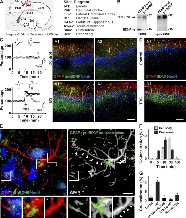
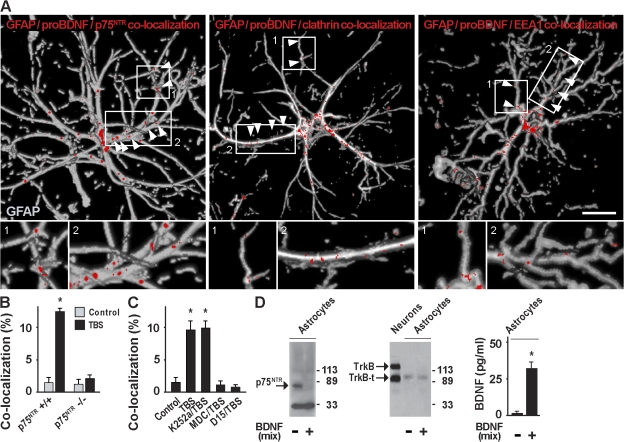
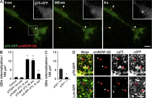
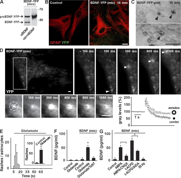
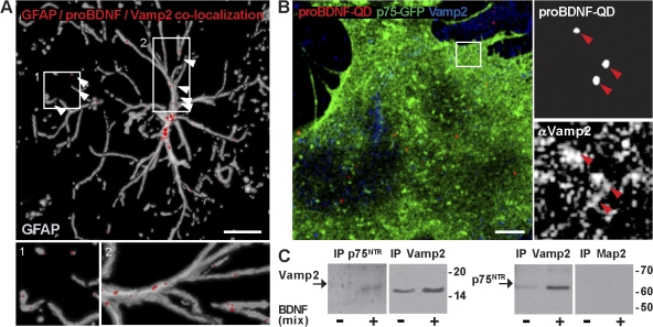
Activity-dependent secretion of brain-derived neurotrophic factor (BDNF) is thought to enhance synaptic plasticity, but the mechanisms controlling extracellular availability and clearance of secreted BDNF are poorly understood. We show that BDNF is secreted in its precursor form (pro-BDNF) and is then cleared from the extracellular space through rapid uptake by nearby astrocytes after theta-burst stimulation in layer II/III of cortical slices, a paradigm resulting in long-term potentiation of synaptic transmission. Internalization of pro-BDNF occurs via the formation of a complex with the pan-neurotrophin receptor p75 and subsequent clathrin-dependent endocytosis. Fluorescence-tagged pro-BDNF and real-time total internal reflection fluorescence microscopy in cultured astrocytes is used to monitor single endocytic vesicles in response to the neurotransmitter glutamate. We find that endocytosed pro-BDNF is routed into a fast recycling pathway for subsequent soluble NSF attachment protein receptor-dependent secretion. Thus, astrocytes contain an endocytic compartment competent for pro-BDNF recycling, suggesting a specialized form of bidirectional communication between neurons and glia.


Figures

Figure 1.
Figure 1.
Transfer of pro-BDNF from neurons to perineuronal astrocytes. (A) Schematic representation of the slice preparation. (B) Western blot analysis of BDNF (mix) or cleavage-resistant pro-BDNF (Mowla et al., 2001) using α-BDNF– or α–pro-BDNF–specific antibodies. (C) Field potential amplitudes (black circles) and BDNF levels (gray circles) upon basal (control) or TBS stimulations. After recording, slices were immunostained using α–pro-BDNF. Immunoreactivity is shown in two adjacent areas corresponding to areas A1 and A2 of A. (D) Immunohistochemistry using α-BDNF. Bars, 100 μm. (E) High resolution confocal images of A1 in a slice 20 min after TBS. Pro-BDNF immunoreactivity is shown at the site of astrocytic contact with a neuron (box and inset 1), the astrocytic cell body (box and inset 2), and processes (box and inset 3). Colocalization of pro-BDNF with GFAP immunoreactivity is shown and superimposed onto the 3D reconstruction of the GFAP signal. Arrowheads indicate pro-BDNF immunoreactive puncta distributed along the astrocytic processes. Bar, 20 μm. (F) Time course of pro-BDNF/GFAP colocalization (four slices and nine cells). (G) Pro-BDNF/GFAP colocalization in astrocytes of control (three slices and 12 cells) or TBS slices in the absence (six slices and 24 cells) or presence of anisomycin (five slices and 11 cells), TrkB-Fc (four slices and nine cells), and plasmin (five slices and 24 cells) 10 min after stimulation. NeuN, neuronal nuclei. Data are means ± SEM (error bars). *, P ≤ 0.05.
Figure 2.
Figure 2.
p75NTR–clathrin-mediated internalization of pro-BDNF in astrocytes. (A) Colocalization between GFAP, pro-BDNF and p75NTR, clathrin, or EEA1 immunoreactivity in astrocytes 10 min after slice exposure to TBS. Colocalization signal is shown at the site of astrocytic contact with a neuron (box and inset 1) and astrocytic processes (box and inset 2). Bar, 10 μm. (B) Pro-BDNF/GFAP colocalization in astrocytes of control (five slices and 22 cells) or TBS slices (five slices and 18 cells) in p75NTR+/+ and p75NTR−/− mice. (C) Pro-BDNF/GFAP colocalization in astrocytes of control (six slices and nine cells) or TBS slices in the absence (four slices and 12 cells) or presence of K252a (four slices and nine cells), MDC (three slices and 11 cells), or D15 (three slices and 11 cells). (D) Western blot showing surface expression of p75NTR, TrkB, or TrkB-t from control astrocytes or astrocytes exposed to BDNF (mix). TrkB and TrkB-t expression from cultured neurons is shown for comparison. The right panel shows ELISA measurement of BDNF concentration in astrocytes. Data are means ± SEM (error bars). *, P ≤ 0.05.
Figure 3.
Figure 3.
Internalization of the pro-BDNF–p75NTR complex in cultured astrocytes. (A) Time sequence of pro-BDNF–QD–p75-GFP internalization in cultured astrocytes by TIRF imaging. White arrowheads indicate pro-BDNF–QDs (red) in close proximity to the membrane of an astrocyte transfected with p75-GFP (green). Yellow arrowheads point to reference QDs. Insets depict p75-GFP fluorescence that concentrates at the site of the QD. Bar, 5 μm. (B) Pro-BDNF–QD internalization in astrocytes transfected with p75-GFP (nine cells) or Lck-GFP (six cells). (C) Pro-BDNF–QD internalization in astrocytes from p75NTR+/+ (22 cells) and p75NTR−/− (11 cells) mice. (D) Immunocytochemistry showing colocalization (arrowheads) between QDs (blue) and p75NTR (red) in astrocytes transfected with p75-GFP or Lck-GFP (green). Bar, 2 μm. Data are means ± SEM (error bars). *, P ≤ 0.05.
Figure 4.
Figure 4.
Astrocytes recycle endocytic pro-BDNF for regulated secretion. (A) Western blot analysis of BDNF-YFP (mix) using α-BDNF or α–pro-BDNF antibodies. (B) Immunocytochemistry in astrocytes untreated (n = 12) or incubated for 10 min with BDNF-YFP (mix; n = 18) followed by acid strip. Bar, 10 μm. (C) Ultrastructural characterization of astrocytes exposed to BDNF-YFP gold for 10 min. Arrowheads point to gold particles contained in vesicular organelles. Bar, 500 nm. (D) Representative TIRF images of astrocytes incubated with BDNF-YFP for 5 min. The top sequence depicts exocytic fusion (arrowheads) in a selected astrocytic area (white rectangle) before and after perfusion with glutamate. The bottom sequence shows fusion of a single vesicle. Fluorescence intensity is measured in a circular mask centered over the vesicle and in a concentric annulus on the circle. Bars, 2 μm. (E) Time distribution of fusion events (flashes) after glutamate application. The inset shows the total number of flashes per astrocyte before (n = 17) and after (n = 13) glutamate. (F) ELISA quantification of BDNF secretion from astrocytes before (n = 18) and after (n = 22) glutamate application for 5 min. Astrocytes previously exposed to BDNF (mix) for 10 min were stimulated with glutamate in the absence (n = 14) or presence (n = 4) of TeNT. (G) Secretion of BDNF induced by AMPA or t-ACPD in the absence (n = 23) or presence (n = 17) of the respective antagonists CNQX or AIDA and by 50 Hz (n = 6). Data are means ± SEM (error bars). *, P ≤ 0.05.
Figure 5.
Figure 5.
Vesicles containing pro-BDNF–p75NTR express the Vamp2 component of the SNARE core complex for vesicle fusion. (A) Colocalization between GFAP, pro-BDNF, and Vamp2 immunoreactivity in astrocytes 10 min after TBS. Colocalization signal (arrowheads) is shown at the site of astrocytic contact with a neuron (box and inset 1) and astrocytic processes (box and inset 2). Bar, 20 μm. (B) Immunocytochemistry showing colocalization between pro-BDNF–QDs and Vamp2 in astrocytes transfected with p75-GFP. Right panels depict QD/Vamp2 colocalization (arrowheads) in a selected astrocytic area (boxed area). Bar, 2 μm. (C) Western blot showing p75NTR and Vamp2 expression in endocytic vesicles immunopurified (IP) by magnetic beads coated with α-p75NTR, α-Vamp2, or α-Map2 from astrocytes untreated or treated with BDNF (mix).

References

    1. Aicardi, G., E. Argilli, S. Cappello, S. Santi, M. Riccio, H. Thoenen, and M. Canossa. 2004. Induction of long-term potentiation and depression is reflected by corresponding changes in secretion of endogenous brain-derived neurotrophic factor. Proc. Natl. Acad. Sci. USA. 101:15788–15792. - PMC - PubMed
    1. Allen, N.J., and B.A. Barres. 2005. Signaling between glia and neurons: focus on synaptic plasticity. Curr. Opin. Neurobiol. 15:542–548. - PubMed
    1. An, J.J., K. Gharami, G.Y. Liao, N.H. Woo, A.G. Lau, F. Vanevski, E.R. Torre, K.R. Jones, Y. Feng, B. Lu, and B. Xu. 2008. Distinct role of long 3′ UTR BDNF mRNA in spine morphology and synaptic plasticity in hippocampal neurons. Cell. 134:175–187. - PMC - PubMed
    1. Balkowiec, A., and D.M. Katz. 2000. Activity-dependent release of endogenous brain-derived neurotrophic factor from primary sensory neurons detected by ELISA in situ. J. Neurosci. 20:7417–7423. - PMC - PubMed
    1. Bezzi, P., V. Gundersen, J.L. Galbete, G. Seifert, C. Steinhauser, E. Pilati, and A. Volterra. 2004. Astrocytes contain a vesicular compartment that is competent for regulated exocytosis of glutamate. Nat. Neurosci. 7:613–620. - PubMed

Publication types

MeSH terms

Substances