Review
. 2008 Oct;65(10):1291-5.
doi: 10.1001/archneur.65.10.1291.

Progress in periventricular leukomalacia

Affiliations
Review

Progress in periventricular leukomalacia

Wenbin Deng et al. Arch Neurol. 2008 Oct.

Abstract

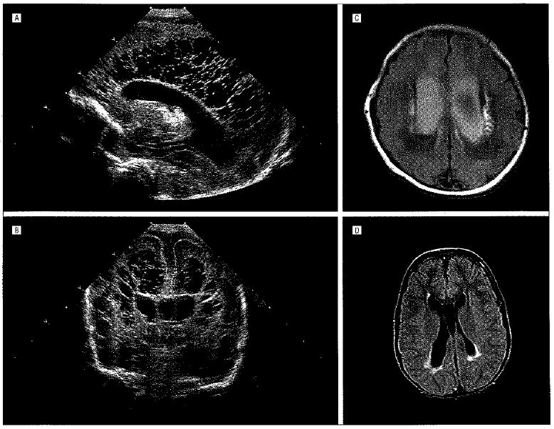
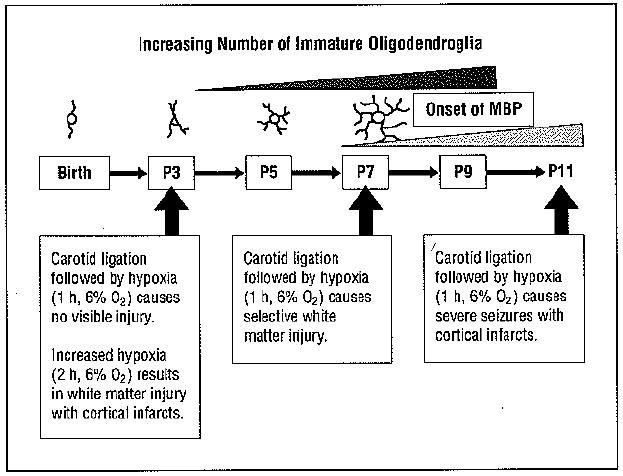
Periventricular leukomalacia (PVL) is the predominant form of brain injury and the leading known cause of cerebral palsy and cognitive deficits in premature infants. The number of low-birth-weight infants who survive to demonstrate these neurologic deficts is increasing. Magnetic resonance imaging-based neuroimaging techniques provide greater diagnostic sensitivity for PVL than does head ultrasonography and often document the involvement of telencephalic gray matter and long tracts in addition to periventricular white matter. The neuropathologic hallmarks of PVL are microglial activation and focal and diffuse periventricular depletion of premyelinating oligodendroglia. Premyelinating oligodendroglia are highly vulnerable to death caused by glutamate, free radicals, and proinflammatory cytokines. Studies in animal models of PVL suggest that pharmacologic interventions that target these toxic molecules will be useful in diminishing the severity of PVL.


Figures

Figure 1
Figure 1
Periventricular leukomalacia (PVL). Sagittal (A) and coronal (B) cranial untrasonograms obtained at age 2 days. Images exemplify extensive bilateral cystic PVL. This twin born at 29 weeks of gestation had fetal heart rate decelerations and Apgar scores of 4 and 7 at 1 and 5 minutes, respectively, related to his role as donor in twin-twin transfusion syndrome. C, Magnetic resonance image (fluid-attenuated inversion recovery) obtained at age 10 days in an infant born at 34 weeks of gestation. Image shows bilateral enlarged, blood-filled ventricles and prominent bilateral periventricular high-signal areas in the white matter lateral to the ventricles, consistent with PVL. The mother, who had diabetes, had pregnancy-induced hypertension, which was treated with magnesium sulfate. The baby, with Apgar scores of 2 and 2 at 1 and 5 minutes, respectively, developed status epilepticus that responded to treatment with phenobarbital sodium. Shortly after birth, he developed severe bilateral intraventricular hemorrhages with posthemorrhagic hydrocephalus, a frequent concomitant of PVL. D, T2-weighted magnetic resonance image (fluid-attenuated inversion recovery) in a 9-year-old girl with intractable complex seizures since infancy. There are peritrigonal high-signal areas, consistent with PVL. She had been born at 37 weeks of gestation to a mother with diabetes, and developed severe hypoxia. which was treated with extracorporeal membrane oxygenation.
Figure 2
Figure 2
Rodent model of periventricuiar leukomalacia. The pattern of hypoxic-ischemic brain injury is both highly age-specific and dependent on the severity of the insult. The complexity of processes elaborated by cells of the oligodendroglial lineage as a function of developmental stage is shown in schematic fashion, ranging from the 1 or 2 processes characteristic of motile oligodendroglial progenitors to the complex process network of premyelinating oligodendroglia. MBP indicates myelin basic protein; P, postnatal day.

References

    1. Back SA, Riddle A, McClure MM. Maturation-dependent vulnerability of perinatal white matter in premature birth. Stroke. 2007;38(2 suppl):724–730. - PubMed
    1. McQuillen PS, Ferreiro DM. Perinatal subplate neuron injury: implications for cortical development and plasticity. Brain Pathol. 2005;15(3):250–260. - PMC - PubMed
    1. De Vries LS, Van Haastert I-LC, Rademaker KJ, Koopman C, Groenendaal F. Ultrasound abnormalities preceding cerebral palsy in high-risk preterm infants. J Pediatr. 2004;144(6):815–820. - PubMed
    1. Inder TE, Huppi PS, Warfield S, et al. Periventricular white matter injury in the premature infant is followed by reduced cerebral cortical gray matter volume at term. Ann Neurol. 1999;46(5):755–760. - PubMed
    1. Nagae LM, Hoon AH, Jr, Stashinko E, et al. Diffusion tensor imaging in children with periventricular leukomalacia: variability of injuries to white matter tracts. AJNR Am J Neuroradiol. 2007;28(7):1213–1222. - PMC - PubMed

Publication types

MeSH terms