. 2008 Oct 28;118(18):1837-47.
doi: 10.1161/CIRCULATIONAHA.108.793869. Epub 2008 Oct 13.

Increased inflammatory gene expression in ABC transporter-deficient macrophages: free cholesterol accumulation, increased signaling via toll-like receptors, and neutrophil infiltration of atherosclerotic lesions

Affiliations

Increased inflammatory gene expression in ABC transporter-deficient macrophages: free cholesterol accumulation, increased signaling via toll-like receptors, and neutrophil infiltration of atherosclerotic lesions

Laurent Yvan-Charvet et al. Circulation. .

Abstract

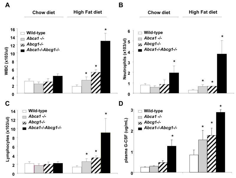
Background: Two macrophage ABC transporters, ABCA1 and ABCG1, have a major role in promoting cholesterol efflux from macrophages. Peritoneal macrophages deficient in ABCA1, ABCG1, or both show enhanced expression of inflammatory and chemokine genes. This study was undertaken to elucidate the mechanisms and consequences of enhanced inflammatory gene expression in ABC transporter-deficient macrophages.

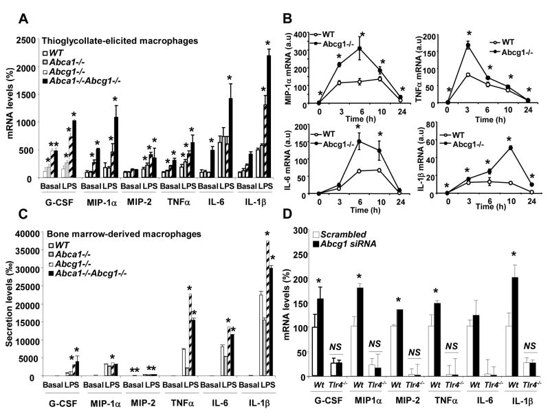
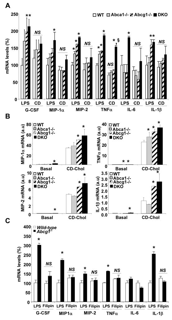
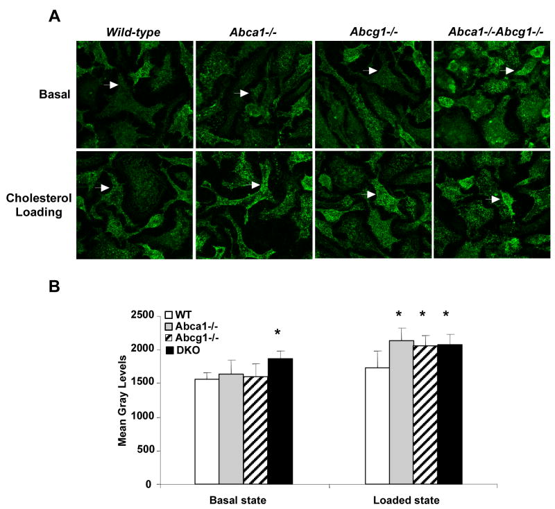
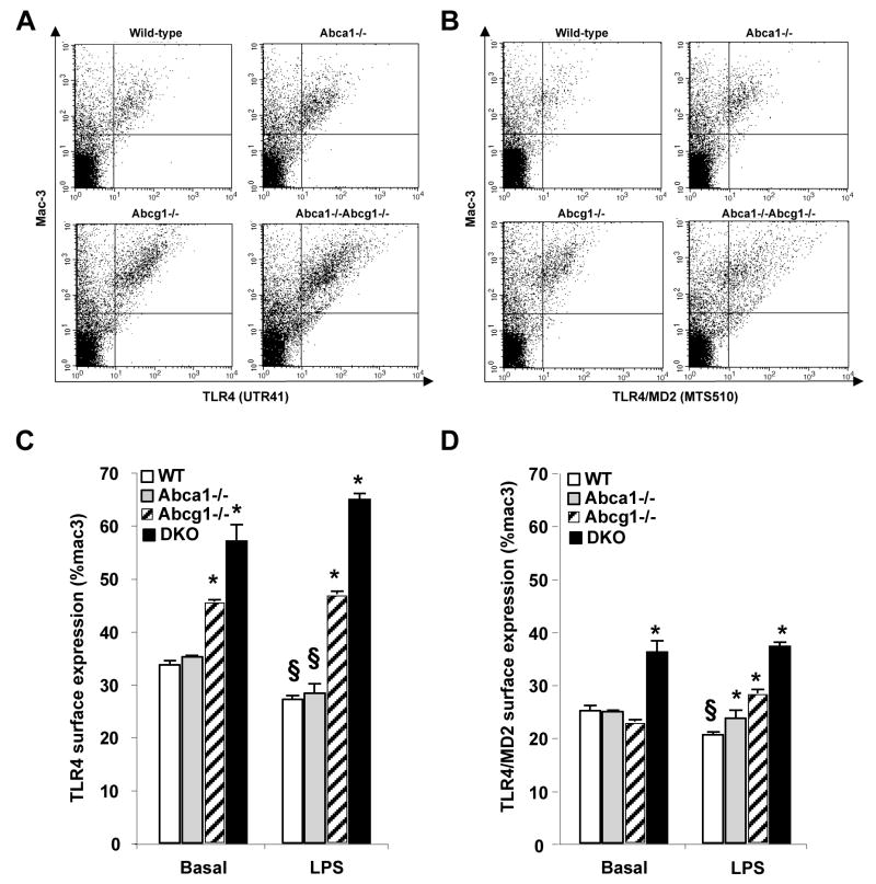
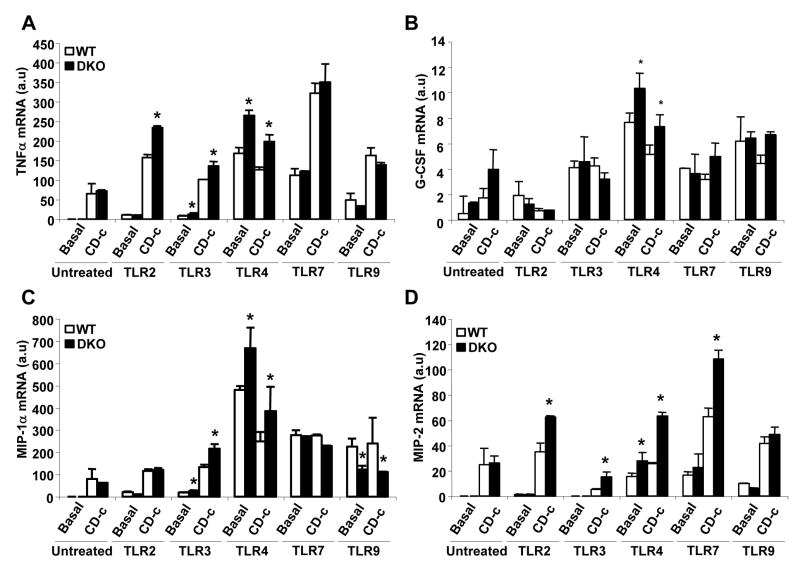
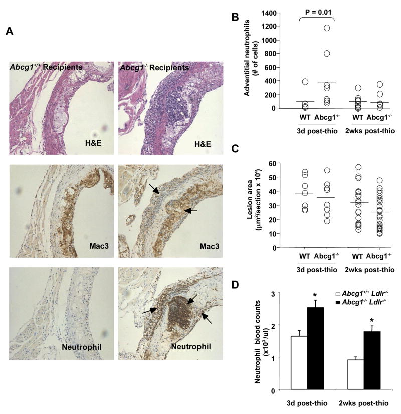
Methods and results: Basal and lipopolysaccharide-stimulated thioglycollate-elicited peritoneal macrophages showed increased inflammatory gene expression in the order Abca1(-/-)Abcg1(-/-)>Abcg1(-/-)>Abca1(-/-)>wild-type. The increased inflammatory gene expression was abolished in macrophages deficient in Toll-like receptor 4 (TLR4) or MyD88/TRIF. TLR4 cell surface concentration was increased in Abca1(-/-)Abcg1(-/-)>Abcg1(-/-)> Abca1(-/-)> wild-type macrophages. Treatment of transporter-deficient cells with cyclodextrin reduced and cholesterol-cyclodextrin loading increased inflammatory gene expression. Abca1(-/-)Abcg1(-) bone marrow-derived macrophages showed enhanced inflammatory gene responses to TLR2, TLR3, and TLR4 ligands. To assess in vivo relevance, we injected intraperitoneally thioglycollate in Abcg1(-/-) bone marrow-transplanted, Western diet-fed, Ldlr-deficient mice. This resulted in a profound inflammatory infiltrate in the adventitia and necrotic core region of atherosclerotic lesions, consisting primarily of neutrophils.

Conclusions: The results suggest that high-density lipoprotein and apolipoprotein A-1 exert anti-inflammatory effects by promoting cholesterol efflux via ABCG1 and ABCA1 with consequent attenuation of signaling via Toll-like receptors. In response to a peripheral inflammatory stimulus, atherosclerotic lesions containing Abcg1(-/-) macrophages experience an inflammatory "echo," suggesting a possible mechanism of plaque destabilization in subjects with low high-density lipoprotein levels.


Conflict of interest statement

Disclosures: Conflict of interest, Alan R. Tall reports being a consultant to Pfizer, Merck, Boehringer-Ingelheim and Takeda Pharmaceuticals.

Figures

Figure 1
Figure 1. Enhanced inflammatory and chemokine gene expression in Abca1−/−, Abcg1−/− and Abca1−/−Abcg1−/− compared to WT macrophages in response to LPS
Thioglycollate-elicited or bone marrow-derived macrophages from mice on mixed background C57BL/6 × DBA were incubated in 0.2% BSA DMEM in the presence of 50ng/mL LPS for 4h (A and C, respectively). C57BL/6 WT and Abcg1−/− thioglycollate-elicited macrophages were incubated for various times with 50ng/mL LPS (B). Thioglycollate-elicited macrophages from WT and Tlr4−/− mice were treated with scrambled or ABCG1 siRNA before LPS stimulation for 4h (D). At the end of the incubation, transcript or secretion levels were determined. Expression of mRNA was normalized to β-actin. mRNA and secretion levels were expressed as percentage over untreated WT macrophages. Results are mean ± SEM of three independent experiments. *P< 0.05 vs. WT.
Figure 2
Figure 2. Modulation of the inflammatory response in Abca1−/−, Abcg1−/− and Abca1−/− Abcg1−/− macrophages after manipulation of plasma membrane cholesterol
Thioglycollate-elicited macrophages from mice on mixed background C57BL/6 × DBA were incubated with 5mM cyclodextrin (CD) for 30 min before treatment with 50ng/mL LPS for 4h (A) or incubated with CD-cholesterol (2.5:1 molar ratio) for 4 hours (B). Filipin was pre-incubated at 3μg/mL for 30 min before addition of LPS to the cells and during the 4 hours-LPS treatment (C). Inflammatory transcript levels were quantified and normalized to β-actin RNA amount. mRNA levels were expressed as percentage over LPS-treated WT macrophages or as arbitrary units (a.u). Values are mean ± SEM. *P< 0.05 vs. WT.
Figure 3
Figure 3. Visualized lipid rafts in Abca1−/−, Abcg1−/− and Abca1−/−Abcg1−/− macrophages
Bone marrow-derived macrophages from mice on mixed background C57BL/6 × DBA were cultured in 0.2% BSA DMEM (Basal) or loaded overnight with 50μg/mL acLDL plus 3μmol/L TO901317 compound (Loaded state). Plasma membrane raft structures were labeled for ganglioside GM1 and visualized by confocal microscopy as described in methods. Images represent a 3D re-construction from the z-stack of image slices (A). Quantification was performed using ImageJ software and expressed as mean gray levels (B). Values are mean ± SD. *P< 0.05 vs. WT.
Figure 4
Figure 4. Increased surface expression of TLR4/MD2 complex in Abca1−/−, Abcg1−/−and Abca1−/−Abcg1−/− macrophages
Thioglycollate-elicited macrophages from mice of each genotype were were cultured for 1h in suspension in presence or absence of 50ng/mL LPS. Cell surface expression of TLR4 (A and C) or TLR4/MD2 complex (B and D) was detected by flow cytometry of live cells stained with Alexa Fluor 488 anti-TLR4 (UT41), FITC anti-mouse TLR4/MD2 complex (MTS510) and PE anti-mouse Mac-3 staining (A and D). Profiles of fluorescence intensity of TLR4, TLR4/MD2 complex and Mac-3 staining in LPS-treated condition (A and B, respectively). Change in TLR4 or TLR4/MD2 surface expression in the different conditions (C and D, respectively). Data are mean ± SEM and expressed as % of Mac-3. *P< 0.05 vs. WT cells in the same condition. §P< 0.05 vs. untreated condition.
Figure 5
Figure 5. Increased response to TLR2, TLR3 and TLR4 ligands in Abca1−/−, Abcg1−/−and Abca1−/−Abcg1−/− macrophages
Bone marrow-derived macrophages from mice on mixed background C57BL/6 × DBA were incubated in 10% FBS DMEM (basal) or with CD-cholesterol (CD-c, 2.5:1 molar ratio) for 4 hours in presence of different TLR ligands: TLR2 ligand (peptidoglycan, PGN, 2.5μg/mL), TLR3 ligand (PolyI:C, 2.5μg/mL), TLR4 ligand (LipidA, 100ng/mL), TLR7 ligand (Gardiquimod, 2.5μg/mL) and TLR9 (Bacterial CpG-DNA, 2.5μg/mL). Inflammatory transcript levels were quantified and normalized to β-actin RNA amount. mRNA levels of TNFα (A), G-CSF (B), MIP-1α (C) and MIP-2 (D) were expressed as arbitrary units (a.u). Values are mean ± SEM. *P< 0.05 vs. WT.
Figure 6
Figure 6. Increased neutrophil infiltration in the proximal aorta of Ldlr−/− mice transplanted with Abcg1−/− bone marrow but no significant change in atherosclerotic lesion development following an acute inflammatory stimulus
H&E staining in the proximal aorta of C57BL/6 Abcg1−/− recipients challenged for 3 days with a single i.p injection of thioglycollate (A). Mac-3 and MCA771G immunostaining (brown) revealed typical accumulations of foam cell macrophages under fibrous caps as well as prominent accumulations of neutrophils in the adventicia underlying plaques, smaller numbers of cells underneath fibrous caps, and neutrophil debris within necrotic cores (A). Quantification of the neutrophil infiltration in aortas. The increased neutrophil infiltration observed in Abcg1−/− recipients 3 days after thioglycollate injection was transient since no significant differences were observed 2 weeks post-injection (B). Quantification of proximal aortic root lesion area in wild-type and Abcg1−/− recipients revealed no apparent effect of the neutrophil infiltration on lesion areas (C). Increased peripheral blood neutrophil counts in Abcg1−/− recipients may contribute to increased neutrophil infiltration of the aortic root (D). *P<0.05 vs. WT recipients.
Figure 7
Figure 7. Increased peripheral blood neutrophils and plasma G-CSF in Abca1−/−, Abcg1−/− and Abca1−/−Abcg1−/− total knockout female mice fed a high fat diet for 11 weeks
Peripheral white blood count, WBC (A), blood neutrophil (B), and lymphocyte counts (C). Increased plasma G-CSF levels closely paralleled the neutrophilia observed in mice carrying the different genotypes (D). Data are mean ± SEM. *P<0.05 vs. WT

Similar articles

Cited by

References

    1. Gordon DJ, Rifkind BM. High-density lipoprotein-the clinical implications of recent studies. N Engl J Med. 1989;321:1311–1316. - PubMed
    1. Plump AS, Scott CJ, Breslow JL. Human apolipoprotein A–I gene expression increases high density lipoprotein and suppresses atherosclerosis in the apolipoprotein E-deficient mouse. Proc Natl Acad Sci USA. 1994;91:9607–9611. - PMC - PubMed
    1. Okamoto H, Yonemori F, Wakitani K, Minowa T, Maeda K, Shinkai H. A cholesteryl ester transfer protein inhibitor attenuates atherosclerosis in rabbits. Nature. 2000;406:203–207. - PubMed
    1. Kontush A, Chapman MJ. Functionally defective high-density lipoprotein: a new therapeutic target at the crossroads of dyslipidemia, inflammation, and atherosclerosis. Pharmacol Rev. 2006;58:342–374. - PubMed
    1. Oram JF, Lawn RM, Garvin MR, Wade DP. ABCA1 is the cAMP- inducible apolipoprotein receptor that mediates cholesterol secretion from macrophages. J Biol Chem. 2000;275:34508–34511. - PubMed

Publication types

MeSH terms