. 2008 Oct 28;118(18):1802-9.
doi: 10.1161/CIRCULATIONAHA.108.785881. Epub 2008 Oct 13.

Real-time catheter molecular sensing of inflammation in proteolytically active atherosclerosis

Affiliations

Real-time catheter molecular sensing of inflammation in proteolytically active atherosclerosis

Farouc A Jaffer et al. Circulation. .

Abstract

Background: To enable intravascular detection of inflammation in atherosclerosis, we developed a near-infrared fluorescence (NIRF) catheter-based strategy to sense cysteine protease activity during vascular catheterization.

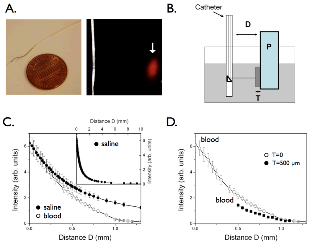
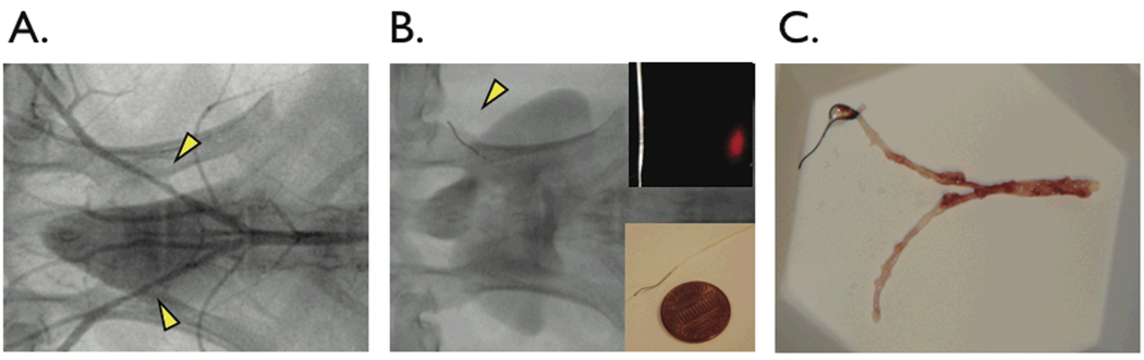
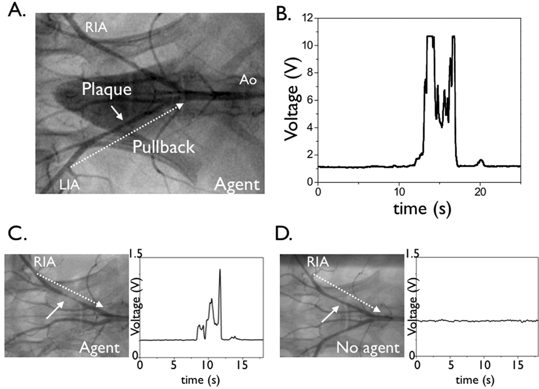
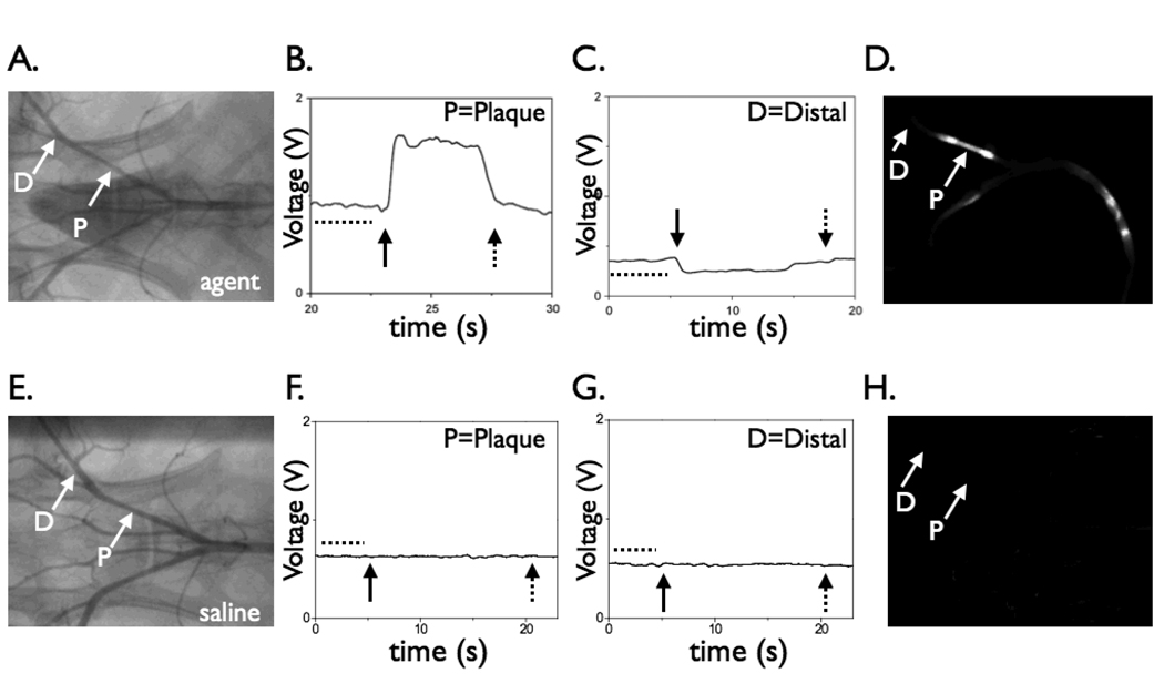
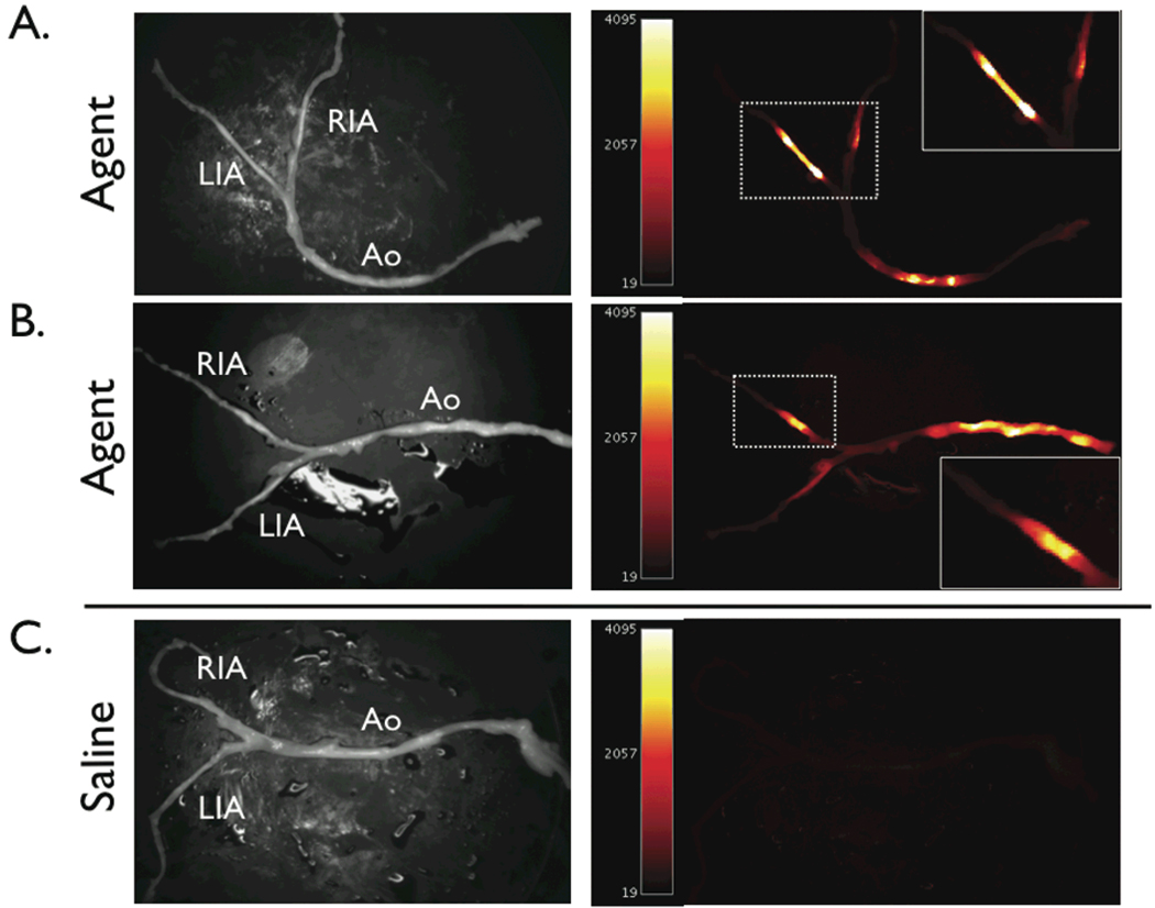
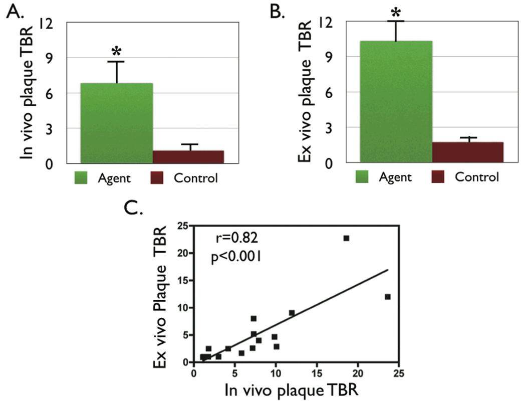
Methods and results: The NIRF catheter design was based on a clinical coronary artery guidewire. In phantom studies of NIRF plaques, blood produced only a mild (<30%) attenuation of the fluorescence signal compared with saline, affirming the favorable optical properties of the NIR window. Catheter evaluation in vivo used atherosclerotic rabbits (n=11). Rabbits received an injection of a cysteine protease-activatable NIRF imaging agent (Prosense750; excitation/emission, 750/770 nm) or saline. Catheter pullbacks through the blood-filled iliac artery detected NIRF signals 24 hours after injection of the probe. In the protease agent group, the in vivo peak plaque target-to-

Background: <0.05). Ex vivo fluorescence reflectance imaging corroborated these results (target-to-

Background: <0.01). In the protease group only, saline flush-modulated NIRF signal profiles further distinguished atheromata from normal segments in vivo (P<0.01). Good correlation between the in vivo and ex vivo plaque target-to-

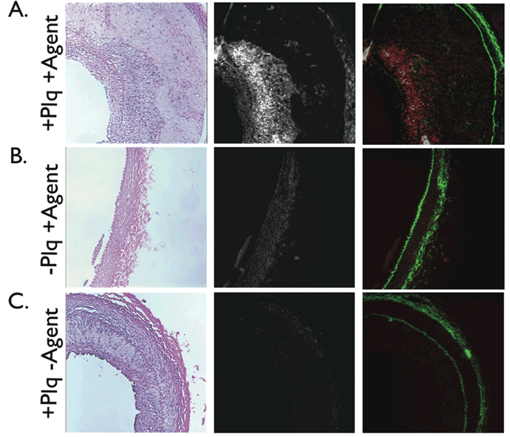
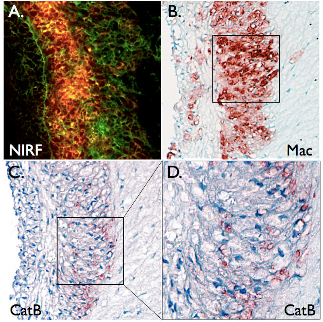
Background: =0.82, P<0.01). Histopathological analyses demonstrated strong NIRF signal in plaques only from the protease agent group. NIRF signals colocalized with immunoreactive macrophages and the cysteine protease cathepsin B.

Conclusions: An intravascular fluorescence catheter can detect cysteine protease activity in vessels the size of human coronary arteries in real time with an activatable NIRF agent. This strategy could aid in the detection of inflammation and high-risk plaques in small arteries.


Figures

Figure 1
Figure 1
The near-infrared fluorescence (NIRF) catheter protoype. The catheter consists of a 0.36 mm/0.014 inch floppy radio-opaque tip with maximum outer diameter 0.48mm/0.019 inches. The focal spot for the 90-degree arc sensing catheter was 40±15 micrometer at a working distance of 2±1 mm (arrow). (B) Phantom for intravascular simulation measurements: P=plaque, consisting of intralipid + India Ink 50 + AF750 (NIR fluorochrome); T=tissue (fibrous cap), consisting of polyester casting resin + titanium dioxide (TiO2) + india ink; the container (grey shaded area) was filled with fresh rabbit blood or saline. The catheter was immersed in fresh rabbit blood and positioned at a variable distance D from a fluorescent phantom representing the plaque P. In order to mimic the presence of a fibrous cap, a solid tissue phantom of thickness T was interposed between the plaque and the lumen. (C) Plot of the detected NIR fluorescence signal as a function of the distance D, in the presence of blood (1/e signal decay 500 micrometers, empty circles) compared to saline (1/e signal decay 700 micrometers, solid circles). The inset shows the fluorescence signal decay in saline at distances up to 10mm. (D) Plot of the detected NIR fluorescence signal in blood in the presence of a tissue phantom T of thickness 500 micrometers shows modest NIRF signal attenuation (<35%) compared to the case in figure 1C where T=0.
Figure 2
Figure 2
In vivo catheter placement in experimental atherosclerosis of New Zealand white rabbits. (A) Angiography of balloon-injured, cholesterol-fed rabbits revealed visible lesions in the iliac arteries (arrowheads). (B) The NIRF catheter guidewire was easily delivered past stenoses in the iliac arteries (arrowhead). (C) Gross pathology revealed yellow-white atheromata in injured areas in the iliac arteries.
Figure 3
Figure 3
Real-time, in vivo fluorescence sensing of inflammation in atherosclerotic vessels. (A) Repeated manual pullback of the catheter was performed in each iliac distal-to-proximal over 20 seconds (dotted arrow, also see Video). (B,C) In rabbits that received the NIRF protease-activatable agent 24 hours beforehand (active group), strong NIRF signal (average TBR 6.8) was detected as the catheter pulled back across the plaque defined by the angiogram. Catheters were re-advanced to lesions and stationary recordings confirmed augmented NIRF signal. (D) In control saline-injected rabbits, minimal NIRF signal deviation was detected during catheter pullback. RIA=right iliac artery, LIA=left iliac artery, Ao=aorta.
Figure 4
Figure 4
Effect of blood absorption on catheter-detected NIRF signals. In vivo NIRF signal profiles of protease-agent and control rabbits under conditions of blood displacement (via balloon occlusion and saline flushing). The NIRF catheter was positioned adjacent to a plaque or distal reference segment. (A) Angiogram of a rabbit receiving the protease agent (“active”) revealing bilateral iliac artery plaques. (B) With the catheter positioned at the right iliac artery plaque, blood displacement via saline flushing augmented the peak NIRF signal (average 74%), as predicted by the phantom studies. (C) In contrast, in normal vessel areas, saline flushing reduced the NIRF signal by 38% (p<0.01), consistent with the displacement of the low-level background fluorescence produced by the circulating, unactivated imaging agent. (D) Corresponding ex vivo fluorescence reflectance image demonstrating strong NIRF signal in the plaque but not distal reference segment. (E) Angiogram of a control animal showing bilateral iliac artery plaques. (F,G) In control animals, blood displacement did not alter the NIRF signal in areas of plaques or normal segments. (H) Negligible NIRF signal was evident on corresponding ex vivo FRI. Images in (D) and (H) windowed identically. In addition, the NIRF signal remained stable during acquisition through physiological blood flow, in both Prosense-injected and control animals, and in both areas of plaques and distal reference segments (dotted line region prior to balloon inflation, figures B, C, F, and G). For (B,C,F,G): closed arrow, start of balloon occlusion and saline flushing; dotted arrow, deflation of the balloon. P=plaque, D=distal.
Figure 5
Figure 5
Ex vivo paired white light and near-infrared fluorescence reflectance images (FRI) of atherosclerotic arteries. (A,B) Augmented NIRF signal was evident in plaques in rabbits injected with the protease-activatable agent (active group). (C) In contrast, control animals showed only minimal autofluorescence signal. NIRF images were windowed equally. RIA=right iliac artery, LIA=left iliac artery, Ao=aorta.
Figure 6
Figure 6
Augmented NIRF signal in atheromata in the protease-agent group. (A) The in vivo peak plaque target-to-background ratio (TBR) was 558% greater in the protease agent group vs. saline (6.8±1.9 vs. 1.3±0.27 respectively, p<0.05). (B) The peak plaque TBR on ex vivo fluorescence reflectance imaging was 856% greater in the protease agent group, with TBR 10.3±1.8 agent vs. 1.8±0.3 saline, p<0.01. (C) Correlation between the in vivo and ex vivo plaque TBR was significant (r=0.82, p<0.01).
Figure 7
Figure 7
Correlative histopathology and fluorescence microscopy of representative arterial sections. Left, H&E ×100; middle, NIR fluorescence microscopy ×100; right, two-channel merged NIR fluorescence (red) and elastin autofluorescence (green) ×100. (A) In plaque sections from the protease agent group, abundant NIRF signal colocalized with cell-rich areas of plaques, (B) but not in the normal vessel wall. (C) In plaque sections from the control group, negligible NIRF signal was detected, consistent with the in vivo and ex vivo NIRF imaging results. NIRF images windowed identically.
Figure 8
Figure 8
Augmented plaque NIRF signals colocalize with plaque macrophages and the cysteine protease cathepsin B. (A) Two-channel fluorescence microscopy (×200) of plaque section in figure 7A demonstrating abundant NIRF signal (red) overlying autofluorescence (green). (B) Immunoreactive macrophages (Mac, ×200) and (C,D) immunoreactive cathepsin B (CatB, ×200 and ×400) colocalize with the NIRF signal.

References

    1. Hansson GK, Libby P. The immune response in atherosclerosis: a double-edged sword. Nat Rev Immunol. 2006;6:508–519. - PubMed
    1. Jaffer FA, Libby P, Weissleder R. Molecular imaging of cardiovascular disease. Circulation. 2007;116:1052–1061. - PubMed
    1. Sanz J, Fayad ZA. Imaging of atherosclerotic cardiovascular disease. Nature. 2008;451:953–957. - PubMed
    1. Chen J, Tung CH, Mahmood U, Ntziachristos V, Gyurko R, Fishman MC, Huang PL, Weissleder R. In vivo imaging of proteolytic activity in atherosclerosis. Circulation. 2002;105:2766–2771. - PubMed
    1. Deguchi JO, Aikawa M, Tung CH, Aikawa E, Kim DE, Ntziachristos V, Weissleder R, Libby P. Inflammation in atherosclerosis: visualizing matrix metalloproteinase action in macrophages in vivo. Circulation. 2006;114:55–62. - PubMed

Publication types

MeSH terms