

Structural basis for glycyl radical formation by pyruvate formate-lyase activating enzyme
- PMID: 18852451
- PMCID: PMC2571006
- DOI: 10.1073/pnas.0806640105
Structural basis for glycyl radical formation by pyruvate formate-lyase activating enzyme
Abstract
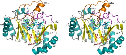
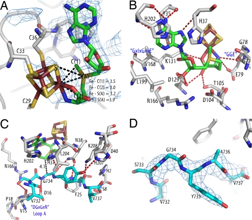
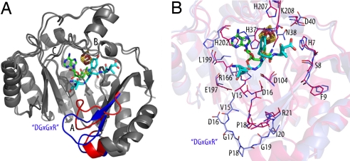
Pyruvate formate-lyase activating enzyme generates a stable and catalytically essential glycyl radical on G(734) of pyruvate formate-lyase via the direct, stereospecific abstraction of a hydrogen atom from pyruvate formate-lyase. The activase performs this remarkable feat by using an iron-sulfur cluster and S-adenosylmethionine (AdoMet), thus placing it among the AdoMet radical superfamily of enzymes. We report here structures of the substrate-free and substrate-bound forms of pyruvate formate-lyase-activating enzyme, the first structures of an AdoMet radical activase. To obtain the substrate-bound structure, we have used a peptide substrate, the 7-mer RVSGYAV, which contains the sequence surrounding G(734). Our structures provide fundamental insights into the interactions between the activase and the G(734) loop of pyruvate formate-lyase and provide a structural basis for direct and stereospecific H atom abstraction from the buried G(734) of pyruvate formate-lyase.
Conflict of interest statement
The authors declare no conflict of interest.
Figures
Similar articles
-
A dehydroalanyl residue can capture the 5'-deoxyadenosyl radical generated from S-adenosylmethionine by pyruvate formate-lyase-activating enzyme.Biochem Biophys Res Commun. 1999 Jan 19;254(2):306-10. doi: 10.1006/bbrc.1998.9931. Biochem Biophys Res Commun. 1999. PMID: 9918833
-
Pyruvate formate-lyase activating enzyme: elucidation of a novel mechanism for glycyl radical formation.Arch Biochem Biophys. 2005 Jan 1;433(1):288-96. doi: 10.1016/j.abb.2004.09.028. Arch Biochem Biophys. 2005. PMID: 15581584 Review.
-
Adenosylmethionine-dependent synthesis of the glycyl radical in pyruvate formate-lyase by abstraction of the glycine C-2 pro-S hydrogen atom. Studies of [2H]glycine-substituted enzyme and peptides homologous to the glycine 734 site.J Biol Chem. 1994 Apr 29;269(17):12432-7. J Biol Chem. 1994. PMID: 8175649
-
Pyruvate formate-lyase and its activation by pyruvate formate-lyase activating enzyme.J Biol Chem. 2014 Feb 28;289(9):5723-9. doi: 10.1074/jbc.M113.496877. Epub 2013 Dec 12. J Biol Chem. 2014. PMID: 24338017 Free PMC article.
-
Glycyl radical activating enzymes: structure, mechanism, and substrate interactions.Arch Biochem Biophys. 2014 Mar 15;546:64-71. doi: 10.1016/j.abb.2014.01.020. Epub 2014 Jan 31. Arch Biochem Biophys. 2014. PMID: 24486374 Free PMC article. Review.
Cited by
-
Computational Approaches: An Underutilized Tool in the Quest to Elucidate Radical SAM Dynamics.Molecules. 2021 Apr 29;26(9):2590. doi: 10.3390/molecules26092590. Molecules. 2021. PMID: 33946806 Free PMC article. Review.
-
Discovery of novel bacterial queuine salvage enzymes and pathways in human pathogens.Proc Natl Acad Sci U S A. 2019 Sep 17;116(38):19126-19135. doi: 10.1073/pnas.1909604116. Epub 2019 Sep 3. Proc Natl Acad Sci U S A. 2019. PMID: 31481610 Free PMC article.
-
The Autonomous Glycyl Radical Protein GrcA Restores Activity to Inactive Full-Length Pyruvate Formate-Lyase In Vivo.J Bacteriol. 2022 May 17;204(5):e0007022. doi: 10.1128/jb.00070-22. Epub 2022 Apr 4. J Bacteriol. 2022. PMID: 35377165 Free PMC article.
-
Solution structure and biochemical characterization of a spare part protein that restores activity to an oxygen-damaged glycyl radical enzyme.J Biol Inorg Chem. 2019 Sep;24(6):817-829. doi: 10.1007/s00775-019-01681-2. Epub 2019 Jun 27. J Biol Inorg Chem. 2019. PMID: 31250200 Free PMC article.
-
A B12-dependent radical SAM enzyme involved in oxetanocin A biosynthesis.Nature. 2017 Apr 20;544(7650):322-326. doi: 10.1038/nature21689. Epub 2017 Mar 27. Nature. 2017. PMID: 28346939 Free PMC article.
References
-
- Sofia HJ, Chen G, Hetzler BG, Reyes-Spindola JF, Miller NE. Radical SAM, a novel protein superfamily linking unresolved steps in familiar biosynthetic pathways with radical mechanisms: Functional characterization using new analysis and information visualization methods. Nucleic Acids Res. 2001;29:1097–1106. - PMC - PubMed
-
- Wang SC, Frey PA. S-Adenosylmethionine as an oxidant: The radical SAM superfamily. Trends Biochem Sci. 2007;32:101–110. - PubMed
-
- Cheek J, Broderick JB. Adenosylmethionine-dependent iron-sulfur enzymes: Versatile clusters in a radical new role. J Biol Inorg Chem. 2001;6:209–226. - PubMed
-
- Blaschkowski HP, Neuer G, Ludwig-Festl M, Knappe J. Routes of flavodoxin and ferredoxin reduction in Escherichia coli: CoA-acylating pyruvate:flavodoxin and NADPH:flavodoxin oxidoreductases participating in the activation of pyruvate formate-lyase. Eur J Biochem. 1982;123:563–569. - PubMed
Publication types
MeSH terms
Substances
Associated data
- Actions
- Actions
Grants and funding
LinkOut - more resources
Full Text Sources
Other Literature Sources
Molecular Biology Databases
Miscellaneous
