. 2008 Oct 21;105(42):16159-64.
doi: 10.1073/pnas.0807486105. Epub 2008 Oct 13.

A Schiff base connectivity switch in sensory rhodopsin signaling

Affiliations

A Schiff base connectivity switch in sensory rhodopsin signaling

Oleg A Sineshchekov et al. Proc Natl Acad Sci U S A. .

Abstract

Sensory rhodopsin I (SRI) in Halobacterium salinarum acts as a receptor for single-quantum attractant and two-quantum repellent phototaxis, transmitting light stimuli via its bound transducer HtrI. Signal-inverting mutations in the SRI-HtrI complex reverse the single-quantum response from attractant to repellent. Fast intramolecular charge movements reported here reveal that the unphotolyzed SRI-HtrI complex exists in two conformational states, which differ by their connection of the retinylidene Schiff base in the SRI photoactive site to inner or outer half-channels. In single-quantum photochemical reactions, the conformer with the Schiff base connected to the cytoplasmic (CP) half-channel generates an attractant signal, whereas the conformer with the Schiff base connected to the extracellular (EC) half-channel generates a repellent signal. In the wild-type complex the conformer equilibrium is poised strongly in favor of that with CP-accessible Schiff base. Signal-inverting mutations shift the equilibrium in favor of the EC-accessible Schiff base form, and suppressor mutations shift the equilibrium back toward the CP-accessible Schiff base form, restoring the wild-type phenotype. Our data show that the sign of the behavioral response directly correlates with the state of the connectivity switch, not with the direction of proton movements or changes in acceptor pK(a). These findings identify a shared fundamental process in the mechanisms of transport and signaling by the rhodopsin family. Furthermore, the effects of mutations in the HtrI subunit of the complex on SRI Schiff base connectivity indicate that the two proteins are tightly coupled to form a single unit that undergoes a concerted conformational transition.


Conflict of interest statement

The authors declare no conflict of interest.

Figures
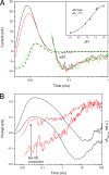
Fig. 1.
Fig. 1.
Laser flash-induced charge movements and absorption changes in free SRI. (A) Photocurrents in wild-type SRI (solid lines) at pH 7.2 (black), 6.9 (red), and 5.4 (green) and in the SRI_D76N mutant at pH 8.0 (dashed black line). Slow region of the signals are duplicated in 20× magnification. (Inset) The pH dependence of maximum current in the wild-type SRI. (B) Charge movement (integral of photocurrent over time) (left axis, black lines) and M intermediate accumulation (absorption changes at 390 nm) (right axis, red lines) in wild-type SRI at pH 7.1 (upper curves) and 6.3 (lower curves). Absorption changes were normalized at their maximum value. Arrows show an increase in the relative amplitude of the fast component of M accumulation corresponding to an increase in the fast outwardly directed charge movement.
Fig. 2.
Fig. 2.
Laser flash-induced charge movements and absorption changes in wild-type SRI–HtrI complex. (A) Charge movements in SRI–HtrI complex (red line) and in free SRI (black line) are compared. (B) Effect of deuteration on the kinetics of charge movements (left axis, solid lines without fitting curves) and M intermediate accumulation (absorption changes at 390 nm) (right axis with dashed fitting curves) in SRI–HtrI complex. Black lines, H2O; red lines, D2O.
Fig. 3.
Fig. 3.
Structural schematic of SRI–HtrI. Shown are locations of amino acid residues that when mutated lead to inverted (marked in red) or recovered (suppressor mutations, marked in green) phototaxis responses. The interaction of SRI and HtrI is schematically presented based on analogy with the SRII–HtrII (40) and HAMP domain (50) atomic structures.
Fig. 4.
Fig. 4.
Laser flash-induced charge movements in inverted and suppressor mutants. (A) Kinetics of charge movements in the inverted (SRI–HtrI_E56Q) (upper red line) and suppressor (SRI_R215W-HtrI_E56Q) (lower black line) mutants. A+ and A, amplitudes of outwardly and inwardly directed proton movements, respectively. (B) Correlation between charge movements and M accumulation in double mutant SR_R215W-HtrI_E56Q. Shown are charge movements (left scale, black lines), and M intermediate accumulation (absorption change at 390 nm) (right scale, red lines) at pH 5.9 (lower curves) and pH 8.0 (upper curves). (C) pH dependence of the ratio of outwardly and inwardly directed proton movements (A+/A) in wild-type SRI–HtrI (black symbols and line) and the inverted SRI–HtrI_E56Q mutant (red symbols and line). (D) Effect of various mutations in receptor and transducer subunits of the SRI–HtrI complex on the ratio of fast outwardly directed charge movement to slower inwardly directed charge movement (A+/A). The ratio values for double suppressor mutants were normalized to corresponding single inverted mutants. The wild-type ratio was normalized to the inverted SRI–HtrI_E56Q mutant.
Fig. 5.
Fig. 5.
Charge movements in D76N mutants. Shown are phenotypically wild-type SRI_D76N-HtrI single mutant (green lines), inverted SRI–HtrI_E56Q single mutant (red lines), and inverted SRI_D76N-HtrI_E56Q double mutant (black lines). Arrow shows the decrease in the amplitude of inwardly directed proton movement.
Fig. 6.
Fig. 6.
Schematic representation. Cartoon depicts light signal transduction by two conformers of the SRI–HtrI complex with opposite Schiff base connectivity assuming switch-coupled channel conformations (see text).

Similar articles

Cited by

References

    1. Ruiz-Gonzalez MX, Marin I. New insights into the evolutionary history of type 1 rhodopsins. J Mol Evol. 2004;58:348–358. - PubMed
    1. Spudich JL, Jung KH. In: Handbook of Photosensory Receptors. Briggs WR, Spudich JL, editors. Weinheim, Germany: Wiley VCH; 2005. pp. 1–21.
    1. Sharma AK, Spudich JL, Doolittle WF. Microbial rhodopsins: Functional versatility and genetic mobility. Trends Microbiol. 2006;14:463–469. - PubMed
    1. Rusch DB, et al. The Sorcerer II Global Ocean Sampling expedition: Northwest Atlantic through eastern tropical Pacific. PLoS Biol. 2007;5:e77. - PMC - PubMed
    1. Spudich JL. The multitalented microbial sensory rhodopsins. Trends Microbiol. 2006;14:480–487. - PubMed

Publication types

LinkOut - more resources