. 2008 Oct 21;105(42):16308-13.
doi: 10.1073/pnas.0806447105. Epub 2008 Oct 13.

Network model of survival signaling in large granular lymphocyte leukemia

Affiliations

Network model of survival signaling in large granular lymphocyte leukemia

Ranran Zhang et al. Proc Natl Acad Sci U S A. .

Abstract

T cell large granular lymphocyte (T-LGL) leukemia features a clonal expansion of antigen-primed, competent, cytotoxic T lymphocytes (CTL). To systematically understand signaling components that determine the survival of CTL in T-LGL leukemia, we constructed a T-LGL survival signaling network by integrating the signaling pathways involved in normal CTL activation and the known deregulations of survival signaling in leukemic T-LGL. This network was subsequently translated into a predictive, discrete, dynamic model. Our model suggests that the persistence of IL-15 and PDGF is sufficient to reproduce all known deregulations in leukemic T-LGL. This finding leads to the following predictions: (i) Inhibiting PDGF signaling induces apoptosis in leukemic T-LGL. (ii) Sphingosine kinase 1 and NFkappaB are essential for the long-term survival of CTL in T-LGL leukemia. (iii) NFkappaB functions downstream of PI3K and prevents apoptosis through maintaining the expression of myeloid cell leukemia sequence 1. (iv) T box expressed in T cells (T-bet) should be constitutively activated concurrently with NFkappaB activation to reproduce the leukemic T-LGL phenotype. We validated these predictions experimentally. Our study provides a model describing the signaling network involved in maintaining the long-term survival of competent CTL in humans. The model will be useful in identifying potential therapeutic targets for T-LGL leukemia and generating long-term competent CTL necessary for tumor and cancer vaccine development.


Conflict of interest statement

The authors declare no conflict of interest.

Figures

Fig. 1.
Fig. 1.
The T-LGL survival signaling network. Node and edge color represents the current knowledge of the signaling abnormalities in T-LGL leukemia. Up-regulated or constitutively active nodes are in red, down-regulated or inhibited nodes are in green, nodes that have been suggested to be deregulated (either up-regulation or down-regulation) are in blue, and the states of white nodes are unknown or unchanged compared with normal. Blue edge indicates activation and red edge indicates inhibition. The shape of the nodes indicates the cellular location: rectangular indicates intracellular components, ellipse indicates extracellular components, and diamond indicates receptors. Conceptual nodes (Stimuli, Cytoskeleton signaling, Proliferation, and Apoptosis) are labeled orange. The full names of the node labels are provided in Table S3.
Fig. 2.
Fig. 2.
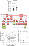
The Boolean model of the T-LGL survival signaling network predicts that constitutive presence of IL-15, and PDGF is sufficient to induce all of the known deregulations in T-LGL leukemia. (A) PDGF-BB is elevated in T-LGL leukemia patient sera compared with normal. Serum level of PDGF-BB from 39 healthy donors (gray triangles) and 22 T-LGL leukemia patients (white diamonds) was assessed by using ELISA. The figure shows a 1.4-fold increase of mean serum level of PDGF-BB (black bar) in T-LGL leukemia patients compared with normal (*, P < 0.005). (B) Hierarchy among known signaling deregulations in T-LGL leukemia. Color code for nodes and edges is the same as in Fig. 1. (C) The effects of IL15, PDGF, and Stimuli on the frequency of apoptosis during simulation. Keeping PDGF ON does not prevent the onset of apoptosis (white triangles). While keeping IL-15 ON, keeping PDGF OFF from the first round of updating delays but cannot prevent the onset of apoptosis (white squares). Setting Stimuli ON at the beginning of the simulation and then keeping it OFF (“ONCE”) does not alter the inhibition of apoptosis upon keeping IL-15 ON in the presence of PDGF (white circles). Results were obtained from 400 simulations of each initial condition. (D) 10 μM AG 1296 specifically induced apoptosis in T-LGL leukemia PBMCs (white circles, n = 4) after 24 h but not in normal PBMCs (gray circles, n = 3, *, P < 0.03). Each circle represents data from one patient or healthy donor. The markers (black bars) indicate the mean apoptosis percentage.
Fig. 3.
Fig. 3.
SPHK1 is a key mediator for the survival of leukemic T-LGL. (A) The effect of SPHK1 inhibition on Apoptosis frequency in the model. The state of SPHK1 was reset to OFF after 15 rounds of updating (white squares), or left unchanged (white diamonds) after a T-LGL-like state was achieved. A rapid increase of apoptosis was observed after SPHK1 inhibition (200 simulations). (B) 20 μM and 40 μM SKI-I selectively induced apoptosis in T-LGL leukemia PBMCs (n = 6) after 48 h but not in normal PBMCs (n = 5, *, P < 0.03 and **, P < 0.01). Each circle represents data from one patient or healthy donor. The markers (black bars) indicate the mean apoptosis percentage. (C) 5 μM and 10 μM SKI-II selectively induced apoptosis in T-LGL leukemia PBMCs after 48 h (white circles, n = 5) but not in normal PBMCs (gray circles, n = 4, *, P < 0.02, and **, P < 0.001). Each circle represents data from one patient or healthy donor. The markers (black bars) indicate the mean apoptosis percentage.
Fig. 4.
Fig. 4.
NFκB is constitutively active in T-LGL leukemia and mediates survival of leukemic T-LGL. (A) Model prediction of the effects of NFκB inhibition (200 simulations). The state of NFκB was reset from ON to OFF after 15 rounds of updating, while keeping IL-15 and PDGF ON. Apoptosis (black squares) was rapidly induced after inhibiting NFκB (black diamonds). The induction of apoptosis was tightly coupled with the down-regulation of Mcl-1 (ж). In contrast, the state of STAT3 (white triangles) remained unchanged until the simulation was terminated. (B) NFκB activity in nuclear extracts of PBMCs from healthy donors and T-LGL leukemia patients. EMSA results are representative of 16 healthy donors and 8 T-LGL leukemia patients tested. White space has been inserted to indicate realigned gel lanes. (C) BAY 11–7082 inhibits NFκB activity in T-LGL leukemia PBMCs. T-LGL leukemia PBMCs were treated with vehicle DMSO or 1 μM, 2 μM, or 5 μM BAY 11–7082 for 3 h, and the activity of NFκB was assessed by EMSA. Result is representative of experiments in three patients. (D) Compared with normal PBMCs (black circles, n = 6), 1 μM BAY 11–7082 selectively induced apoptosis in T-LGL leukemia PBMCs (white circles, n = 6) after 12h treatment (*, P < 0.02). Each circle represents data from one patient or healthy donor. The markers (black bars) indicate the mean of each sample group.
Fig. 5.
Fig. 5.
NFκB-mediated survival pathway in T-LGL leukemia involves PI3K and Mcl-1. (A) Analysis of the potential cause(s) of the constitutive activation of NFκB. As in Table S4, the Boolean logical rule governing the state of NFκB is “NFKB* = [(TPL2 or PI3K) or (FLIP and TRADD and IAP)] and not Apoptosis”. When a T-LGL-like state is achieved, the state of TRADD stabilizes at OFF (see Table S7). Thus, the node that activates NFκB can only be TPL2 or PI3K, which are known to be constitutively active in T-LGL leukemia (see Table S5). Rapid inhibition of NFκB (white squares) was observed after inhibiting PI3K (X) but not after inhibiting TPL2 (gray triangles) (200 simulations). (B) PI3K inhibition induced NFκB inhibition in T-LGL leukemia. T-LGL leukemia PBMCs were treated with vehicle DMSO or 25 μM LY 294002 for 4 h. The amount of total and phospho-AKT was assessed by Western blot assay; NFκB activity was assessed by EMSA. Result is representative of experiments in three patients. (C) NFκB inhibition down-regulates Mcl-1 but does not influence STAT3 activity and the PI3K pathway. T-LGL leukemia PBMCs were treated with vehicle DMSO or 1 μM, 2 μM or 5 μM BAY 11–7082 for 3 h, and the amount of Mcl-1, total- and phospho-AKT was assessed by Western blot assay. STAT3 activity was assessed by EMSA. Result is representative of experiments in three patients. GAPDH was used as a loading control for all of the Western blot assays.
Fig. 6.
Fig. 6.
T-bet is overexpressed and constitutively active in T-LGL leukemia PBMCs. (A) T-bet inhibits IL-2 expression when a T-LGL-like state is achieved. Based on the Boolean logical rule “IL2* = (NFKB or STAT3 or NFAT) and not (TBET or Apoptosis)” (Table S4), T-bet is the only negative regulator of IL-2 expression when cells are still alive. Inhibiting T-bet (white squares) after 15 rounds of updating results in IL-2 (white diamonds) expression after achieving a T-LGL-like state (200 simulations). (B) T-LGL leukemia PBMCs (white squares, n = 10) express 3.3-fold higher T-bet mRNA compared with normal (black circles, n = 5, *, P < 0.02) as assessed by real-time PCR. (C) T-bet protein expression in T-LGL leukemia and normal PBMCs. Western blot assay result is representative of samples from eight healthy donors and six T-LGL leukemia patients. White space has been inserted to indicate realigned gel lanes. (D) T-bet is constitutively active in T-LGL leukemia patients. Nuclear extract from PBMCs of five T-LGL leukemia patients and five healthy donors were tested for their T-bet activity by using EMSA. T-bet exhibited high activity in most T-LGL leukemia patients but not in normal.

References

    1. Klebanoff CA, Gattinoni L, Restifo NP. CD8+ T-cell memory in tumor immunology and immunotherapy. Immunol Rev. 2006;211:214–224. - PMC - PubMed
    1. Sokol L, Loughran TP., Jr Large granular lymphocyte leukemia. Oncologist. 2006;11:263–273. - PubMed
    1. Epling-Burnette PK, et al. Inhibition of STAT3 signaling leads to apoptosis of leukemic large granular lymphocytes and decreased Mcl-1 expression. J Clin Invest. 2001;107:351–362. - PMC - PubMed
    1. Schade AE, Powers JJ, Wlodarski MW, Maciejewski JP. Phosphatidylinositol-3-phosphate kinase pathway activation protects leukemic large granular lymphocytes from undergoing homeostatic apoptosis. Blood. 2006;107:4834–4840. - PMC - PubMed
    1. Epling-Burnette PK, et al. ERK couples chronic survival of NK cells to constitutively activated Ras in lymphoproliferative disease of granular lymphocytes (LDGL) Oncogene. 2004;23:9220–9229. - PubMed

Publication types

MeSH terms

LinkOut - more resources