. 2008;3(10):e3377.
doi: 10.1371/journal.pone.0003377. Epub 2008 Oct 13.

Dendritic cells release HLA-B-associated transcript-3 positive exosomes to regulate natural killer function

Affiliations

Dendritic cells release HLA-B-associated transcript-3 positive exosomes to regulate natural killer function

Venkateswara Rao Simhadri et al. PLoS One. 2008.

Abstract

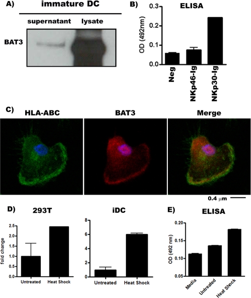
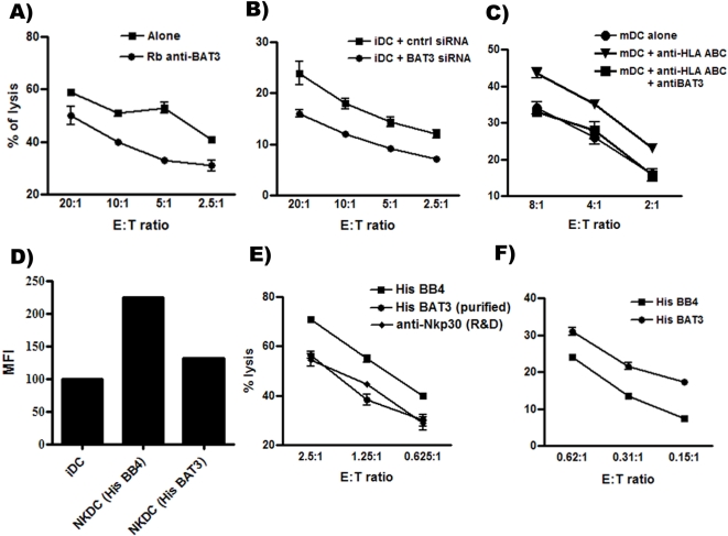
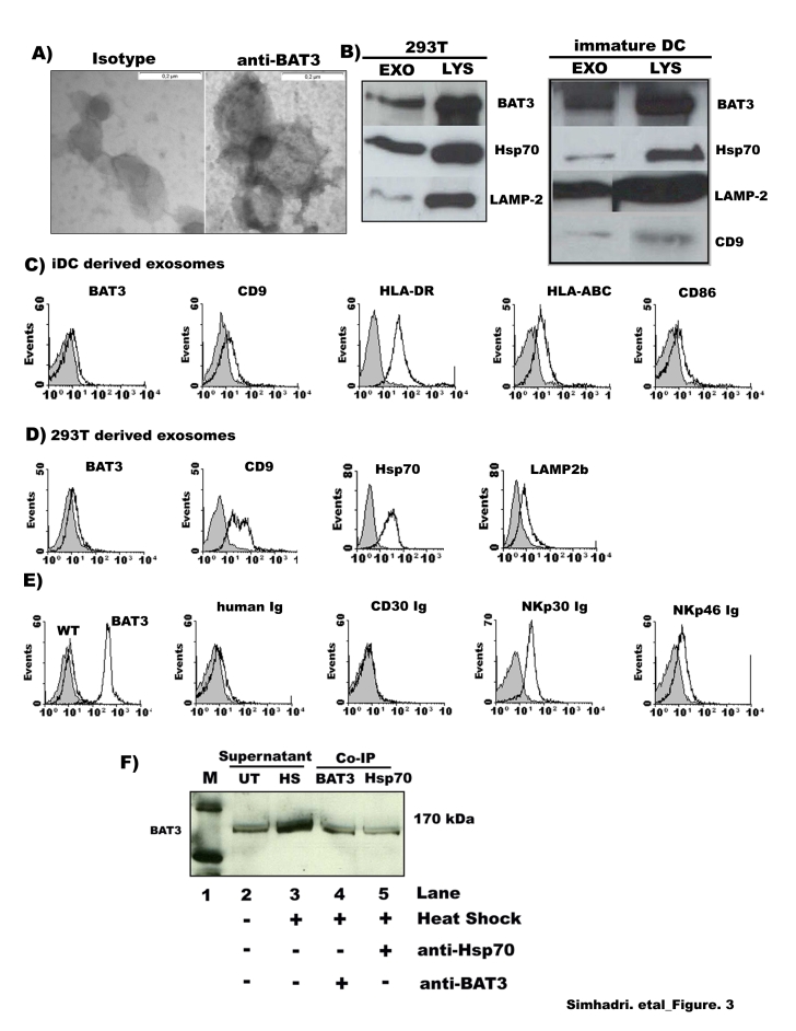
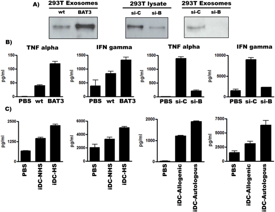
NKp30, a natural cytotoxicity receptor expressed on NK cells is critically involved in direct cytotoxicity against various tumor cells and directs both maturation and selective killing of dendritic cells. Recently the intracellular protein BAT3, which is involved in DNA damage induced apoptosis, was identified as a ligand for NKp30. However, the mechanisms underlying the exposure of the intracellular ligand BAT3 to surface NKp30 and its role in NK-DC cross talk remained elusive. Electron microscopy and flow cytometry demonstrate that exosomes released from 293T cells and iDCs express BAT3 on the surface and are recognized by NKp30-Ig. Overexpression and depletion of BAT3 in 293T cells directly correlates with the exosomal expression level and the activation of NK cell-mediated cytokine release. Furthermore, the NKp30-mediated NK/DC cross talk resulting either in iDC killing or maturation was BAT3-dependent. Taken together this puts forward a new model for the activation of NK cells through intracellular signals that are released via exosomes from accessory cells. The manipulation of the exosomal regulation may offer a novel strategy to induce tumor immunity or inhibit autoimmune diseases caused by NK cell-activation.


Conflict of interest statement

Competing Interests: The authors have declared that no competing interests exist.

Figures

Figure 1
Figure 1. Expression of BAT3 on immature dendritic cells.
(A) Western blot to detect BAT3 in total lysates and supernatant of heat shock treated monocyte-derived iDCs. (B). ELISA plates were coated with recombinant proteins, buffer control (Neg), NKp46-Ig and NKp30-Ig (concentration of 100 ng/ml), followed by incubation with 100 µl of concentrated supernatant obtained from heat shock treated iDCs and detected with anti-BAT3. Data represents absorbance at 492 nm. (C) Laser Scanning Microscopy to visualize HLA- A, B, C and BAT3 on dendritic cells upon staining with specific primary antibodies and labelled secondary antibodies. HLA-A, B, and C (green), BAT3 (red) and merge (right-yellow). Blue represents Hoechst33342 staining of cell nuclei. (D) Quantitative Real time PCR to detect BAT3 mRNA in 293T and iDCs upon exposure to heat shock. The Y-axis determines the fold change; where the untreated samples were normalized to factor 1. (E) Supernatant was collected from iDCs cells either untreated or treated with non-lethal heat shock (Heat shock) and analysed in specific BAT3 ELISA (sandwich method) to determine the amount of BAT3 in the supernatant. Error bars represent the standard deviation of duplicate samples. One representative experiment of three is shown.
Figure 2
Figure 2. Role of BAT3 for iDCs and the effect of purified BAT3 on NK cell function.
(A) Standard Europium release assay: Inhibition of NK-dependent lysis of iDCs in the presence of anti-BAT3 was significant (paired t-test, p-value = 0.008). (B) iDCs transfected with either control siRNA or BAT3 siRNA were co-incubated with NK cells for 4 hours at 37°C. The decrease of iDC lysis upon BAT3 down regulation was significant (p = 0.01). (C) Lysis of mature DCs upon pre-incubation with control antibodies, anti-HLA-ABC and/or anti-BAT3. (D) Co-culture of iDCs with activated NK cells at 5∶1 ratio (iDC∶NK) promotes the maturation of iDCs as shown by FACS analysis to detect expression of the maturation marker CD86. Inhibition of this effect is achieved by soluble purified BAT3. The y-axis represents the mean fluorescence intensity (MFI). (E) The lysis of Raji cells is inhibited by soluble BAT3 and anti-NKp30 compared to the control protein His BB4. The decrease of the lysis was significant (paired t-test, p value = 0.019). (F) NK cells were pre-stimulated with immobilized HisBB4 (control) and purified BAT3 prior a cytotoxicity assay with Raji cells as targets at different effector : target cell ratios. NK cells were derived from different donors for each experiment. Error bars for the lysis experiments represent standard deviation of three replicates. One representative experiment of four is shown.
Figure 3
Figure 3. Bio-chemical characterization of the released BAT3.
(A) Detection of BAT3 expression on exosomes by electron microscopy {left panel: gold antibody control (140000×) and right panel: exosomes stained with anti-BAT3 antibody (140000×)}. (B) Western blotting to detect BAT3, Hsp70, Lamp-2 and CD9 in exosomal fractions (30 µg) and lysate (10 µg) of 293T cells and iDCs. (C,D) FACS analysis to detect BAT3 and various surface markers on exosomes, that were purified from iDCs (C) or 293T cells (D) that were immobilized to latex beads. Grey background represents isotype control. (E) FACS analysis of exosomes derived from control transfected (wt) or BAT3-transfected (BAT3) 293T cells revealed over-expression of BAT3 on the exosomal surface. Specific binding of anti-BAT3, NKp30-Ig and NKp46-Ig was detectable. Grey histograms: background (secondary antibody) staining of beads coated with exosomes. (F) Western blot analysis demonstrates that the enhanced secretion of BAT3 into the supernatant obtained from tumor cells (293T) when treated with heat shock (HS, lane: 3) or left untreated (UT, lane: 2). Lanes 4 and 5 demonstrate the co-immunoprecipitation of BAT3 by using either a polyclonal BAT3 antibody (4th lane) or a monoclonal Ab against Hsp70 (5th lane). The western blot is stained for BAT3. Lane 1 (M) indicates the marker.
Figure 4
Figure 4. Exosomal BAT3 regulates NK cell-function.
(A) Western Blot to detect BAT3 in exosomal fractions upon over expression and depletion. Exosomes purified from untransfected cells (WT), BAT3-transfected cells (BAT3), control siRNA- transfected cells (si-c) and BAT3 siRNA- transfected cells were analysed by Western blotting to detect BAT3. (B) Exosomes were purified from media (PBS), untransfected 293T cells (wt), BAT3-transfected 293T cells (BAT3), control si-RNA (si-c) and BAT3 si-RNA (si-B) transfected 293T cells and used to stimulate NK cells. NK cell-supernatant was collected and used for a cytokine ELISA (TNF-α and IFN-γ). (C) NK cells were stimulated with exosomes derived from untreated iDCs (iDC-NHS exosomes) or upon heat shock (iDC-HS exosomes) for cytokine ELISA (left panels). NK cell-mediated cytokine release was estimated upon stimulation with exosomes derived from allogenic and autologous iDCs (right panels). Primary immune cells were derived from different donors for each experiment. The means of duplicates and the concentration (pg/ml) are indicated. One representative experiment of three is shown.

References

    1. Bryceson YT, Long EO. Line of attack: NK cell specificity and integration of signals. Curr Opin Immunol. 2008;20:344–352. - PMC - PubMed
    1. Bryceson YT, March ME, Ljunggren HG, Long EO. Synergy among receptors on resting NK cells for the activation of natural cytotoxicity and cytokine secretion. Blood. 2006;107:159–166. - PMC - PubMed
    1. Smyth MJ, Cretney E, Kelly JM, Westwood JA, Street SE, et al. Activation of NK cell cytotoxicity. Mol Immunol. 2005;42:501–510. - PubMed
    1. Bloushtain N, Qimron U, Bar-Ilan A, Hershkovitz O, Gazit R, et al. Membrane-associated heparan sulfate proteoglycans are involved in the recognition of cellular targets by NKp30 and NKp46. J Immunol. 2004;173:2392–2401. - PubMed
    1. Hershkovitz O, Jarahian M, Zilka A, Bar-Ilan A, Landau G, et al. Altered glycosylation of recombinant NKp30 hampers binding to heparan sulfate: a lesson for the use of recombinant immunoreceptors as an immunological tool. Glycobiology. 2008;18:28–41. - PubMed

Publication types