. 2008;3(10):e3378.
doi: 10.1371/journal.pone.0003378. Epub 2008 Oct 13.

A new antigen retrieval technique for human brain tissue

Affiliations

A new antigen retrieval technique for human brain tissue

Raúl Alelú-Paz et al. PLoS One. 2008.

Abstract

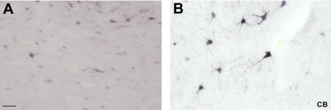
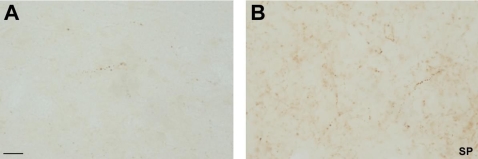
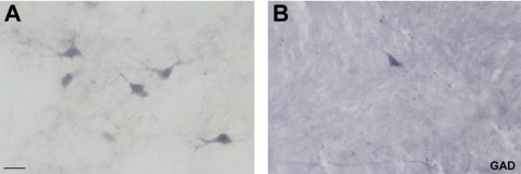
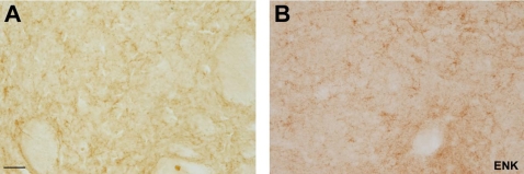
Immunohistochemical staining of tissues is a powerful tool used to delineate the presence or absence of an antigen. During the last 30 years, antigen visualization in human brain tissue has been significantly limited by the masking effect of fixatives. In the present study, we have used a new method for antigen retrieval in formalin-fixed human brain tissue and examined the effectiveness of this protocol to reveal masked antigens in tissues with both short and long formalin fixation times. This new method, which is based on the use of citraconic acid, has not been previously utilized in brain tissue although it has been employed in various other tissues such as tonsil, ovary, skin, lymph node, stomach, breast, colon, lung and thymus. Thus, we reported here a novel method to carry out immunohistochemical studies in free-floating human brain sections. Since fixation of brain tissue specimens in formaldehyde is a commonly method used in brain banks, this new antigen retrieval method could facilitate immunohistochemical studies of brains with prolonged formalin fixation times.


Conflict of interest statement

Competing Interests: The authors have declared that no competing interests exist.

Figures

Figure 1
Figure 1. A, Immunohistochemistry for CB in the MD.
High power view of some CB-ir neurons in the human mediodorsal thalamic nucleus without the employ of citraconic anhydride solution. B, high power view of CB-ir neurons in the same nucleus employing the citraconic anhydride solution. Note the enhancement in the intensity of CB-ir somata. Scale bar: 50 µm.
Figure 2
Figure 2. A, Neuropeptide immunohistochemistry in the human thalamus.
Bright field photomicrographs in high magnification of SP fibers without citraconic anhydride solution in the anteroventral thalamic nucleus. B, bright field photomicrographs in high magnification of SP fibers with citraconic anhydride solution in the same nucleus. Note the enhancement in the intensity of SP-ir fibers and in the number of fibers. Scale bar: 25 µm.
Figure 3
Figure 3. A, Interneuron immunohistochemistry in the MD.
High power view of some GAD-ir neurons in the mediodorsal thalamic nucleus without the use of citraconic anhydride solution. B, high power view of a GAD-ir neuron in the mediodorsal thalamic nucleus utilizing the citraconic anhydride solution. Scale bar: 25 µm.
Figure 4
Figure 4. A–B, Immunohistochemistry for TH in the substantia nigra.
High power view (10× and 20× magnification respectively) of TH-ir neurons in the substantia nigra employing the citraconic anhydride solution. Note the enhancement in the intensity and morphology (somas more preserved and processes more delineated) compared to the conventional immunostaining method (C–D). Scale bar: A and C 100 µm; B and D 50 µm.
Figure 5
Figure 5. A, Immunohistochemistry for ENK in the caudate nucleus.
Bright field photomicrographs in high magnification of ENK fibers without citraconic anhydride solution in the caudate nucleus. B, bright field photomicrographs in high magnification of ENK fibers with citraconic anhydride solution in the same nucleus. Note that the latter fibers are better delineated than in the conventional immunostaing method. Scale bar: 50 µm.
Figure 6
Figure 6. Schematic view of formaldehyde fixation stages.
On the top: formation of methylene glycol by the addition of water and formaldehyde. On the bottom: formaldehyde reacts with various parts of proteins molecules forming adducts. This reaction is followed by the formation of methylene bridges between closed proteins.

Similar articles

Cited by

References

    1. Shi SR, Cote RJ, Taylor CR. Antigen retrieval immunohistochemistry: past, present and future. J Histochem Cytochem. 1997;45:327–343. - PubMed
    1. Mason JT, Ó'Leary TJ. Effects of formaldehyde fixation on protein secondary structure: a calorimetric and infrared spectroscopic investigation. J Histochem Cytochem. 1991;39:225–229. - PubMed
    1. Dapson RW. Macromolecular changes caused by formalin fixation and antigen retrieval. Biotech Histochem. 2007;82:133–40. - PubMed
    1. Shi SR, Key ME, Kalra KL. Antigen retrieval in formalin-fixed, paraffin-embedded tissues: an enhancement method for immunohistochemical staining based on microwave oven heating of tissue sections. J Histochem Cytochem. 1991;39:741–748. - PubMed
    1. Pileri SA, Roncador G, Ceccarelli C, Piccioli M, Briskomatis A, et al. Antigen retrieval techniques in immunohistochemistry: comparison of different methods. J Pathol. 1997;183:116–123. - PubMed

Publication types