. 2008;3(10):e3405.
doi: 10.1371/journal.pone.0003405. Epub 2008 Oct 14.

M-CSF signals through the MAPK/ERK pathway via Sp1 to induce VEGF production and induces angiogenesis in vivo

Affiliations

M-CSF signals through the MAPK/ERK pathway via Sp1 to induce VEGF production and induces angiogenesis in vivo

Jennifer M Curry et al. PLoS One. 2008.

Abstract

Background: M-CSF recruits mononuclear phagocytes which regulate processes such as angiogenesis and metastases in tumors. VEGF is a potent activator of angiogenesis as it promotes endothelial cell proliferation and new blood vessel formation. Previously, we reported that in vitro M-CSF induces the expression of biologically-active VEGF from human monocytes.

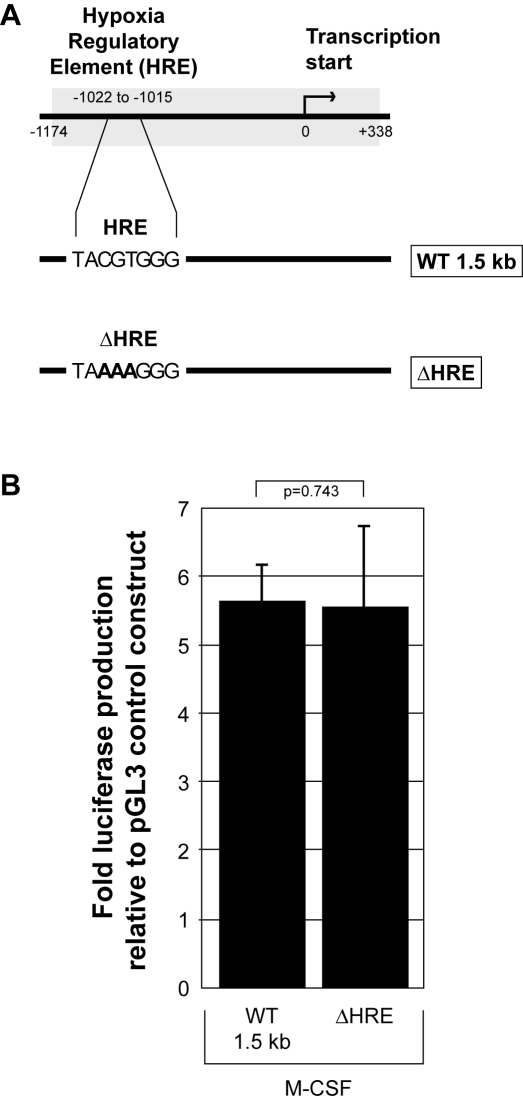
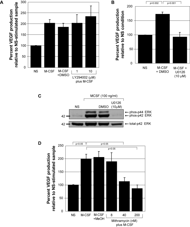
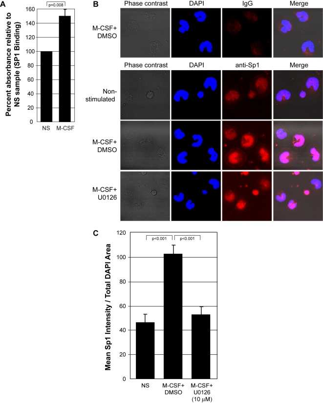
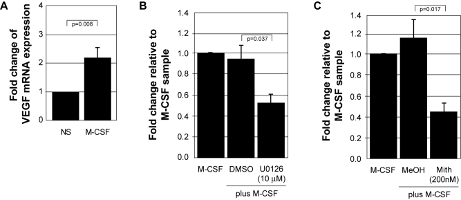
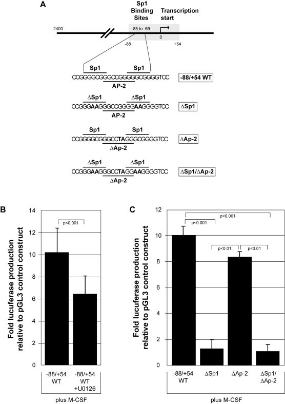
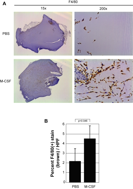
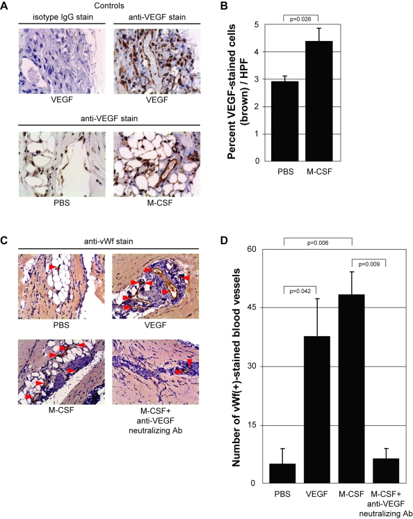
Methodology and results: In this study, we demonstrate the molecular mechanism of M-CSF-induced VEGF production. Using a construct containing the VEGF promoter linked to a luciferase reporter, we found that a mutation reducing HIF binding to the VEGF promoter had no significant effect on luciferase production induced by M-CSF stimulation. Further analysis revealed that M-CSF induced VEGF through the MAPK/ERK signaling pathway via the transcription factor, Sp1. Thus, inhibition of either ERK or Sp1 suppressed M-CSF-induced VEGF at the mRNA and protein level. M-CSF also induced the nuclear localization of Sp1, which was blocked by ERK inhibition. Finally, mutating the Sp1 binding sites within the VEGF promoter or inhibiting ERK decreased VEGF promoter activity in M-CSF-treated human monocytes. To evaluate the biological significance of M-CSF induced VEGF production, we used an in vivo angiogenesis model to illustrate the ability of M-CSF to recruit mononuclear phagocytes, increase VEGF levels, and enhance angiogenesis. Importantly, the addition of a neutralizing VEGF antibody abolished M-CSF-induced blood vessel formation.

Conclusion: These data delineate an ERK- and Sp1-dependent mechanism of M-CSF induced VEGF production and demonstrate for the first time the ability of M-CSF to induce angiogenesis via VEGF in vivo.


Conflict of interest statement

Competing Interests: The authors have declared that no competing interests exist.

Figures

Figure 1
Figure 1. M-CSF induced VEGF production occurs through a HIF-independent mechanism.
A) The 1.5 kb wild type VEGF promoter (WT1.5 kb) and the same promoter sequence containing a non-functional mutation of the hypoxia regulatory element (HRE) (ΔHRE) were inserted into the pGL3-basic vector to create constructs that produce luciferase upon VEGF promoter activation. Nucleotides changed from the original wild type sequence are designated in bold. Human monocytes were transfected with either the control empty construct (pGL3-Basic), or pGL3 containing 1 µg of each of the described VEGF promoter constructs above. B) Transfected monocytes containing (WT1.5 kb) or (ΔHRE) were allowed to adhere for 1 hour in RPMI/5% FBS followed by the addition of fresh media containing rhM-CSF (100 ng/ml) for 16 hours. The adherent cells were lysed and assayed for luciferase production using a luminometer. This data represents the mean+/−SEM of six individual blood donors. All data is represented as the fold change in luciferase over the pGL3 control construct.
Figure 2
Figure 2. M-CSF-induced VEGF production from human monocytes is blocked by ERK inhibition and mithramycin.
A) Monocytes were pre-incubated with DMSO (vehicle control), 1 µM or 10 µM the PI3 kinase inhibitor (LY294002) for 30 minutes. The monocytes were either left non-stimulated (NS), stimulated with rhM-CSF (100 ng/ml) (M-CSF), with rhM-CSF (100 ng/ml) plus DMSO (M-CSF+DMSO), or rhM-CSF (100 ng/ml) plus 1 or 10 µM LY294002 (1) and (10) for 48 hours. Cell-free supernatants were evaluated for VEGF by ELISA. These data represent the mean±SEM from four independent donors. B) Monocytes were pre-incubated with DMSO (vehicle control) or 10 µM of the inhibitor of ERK activity (U0126) for 30 minutes. The monocytes were either left non-stimulated (NS), stimulated with rhM-CSF (100 ng/ml) plus DMSO (M-CSF+DMSO) or with rhM-CSF (100 ng/ml) plus U0126 (M-CSF+U0126 (10 µM)) for 48 hours. Cell-free supernatants were evaluated for VEGF by ELISA. These data represent the mean±SEM from six independent donors. C) Freshly-isolated human monocytes were plated overnight in 5% FBS and M-CSF (20 ng/ml). The next day the cells were starved for 2 hours in minimal media. For the last 30 minutes, DMSO (vehicle control) or 10 µM U0126 was added to the appropriate samples. The cells were left non-stimulated (NS), stimulated with rhM-CSF (100 ng/ml) (M-CSF+DMSO) or (M-CSF+U0126) for 10 minutes. Cell lysates were probed for phospho-ERK MAP Kinase (Thr202) and total ERK MAP Kinase by western blot analysis. This data is representative of two independent monocyte donors. D) Monocytes were pre-incubated with methanol (vehicle control) or 8, 40, or 200 nM of the Sp1 transcription factor binding inhibitor, mithramycin, for 45 minutes. The cells were either left non-stimulated (NS) or stimulated with rhM-CSF (100 ng/ml) (M-CSF), (M-CSF+MeOH), (M-CSF plus 8 nM, 40 nM, or 200 nM mithramycin) for 48 hours. Cell-free supernatants were evaluated for VEGF by ELISA. These data represent the mean±SEM from ten independent blood donors.
Figure 3
Figure 3. M-CSF induces Sp1 nuclear localization in an ERK-dependent manner.
A) Human monocytes were left non-stimulated (NS) or stimulated with rhM-CSF (100 ng/ml) (M-CSF) for 30 minutes. Nuclear lysates were isolated and normalized for total protein. Sp1 that translocated into the nucleus in response to M-CSF was analyzed using a biotinylated Sp1 DNA sequence bound to a streptavidin-coated plate, a polyclonal rabbit anti-Sp1 primary antibody, a HRP-conjugated goat anti-rabbit IgG secondary antibody, and TMB substrate. The absorbance was read at 450 nm to reflect Sp1 within the nucleus. These data represent the mean±SEM from four independent blood donors. B) Human monocytes were starved for 6 hours, inhibited for 30 minutes with U0126 (10 µM) or DMSO (vehicle control) and left non-stimulated (Non-stimulated) or treated with rhM-CSF for 6 hours (M-CSF+DMSO) and (M-CSF+U0126). The cells were fixed, permeablized, and stained with a normal IgG control antibody (top row) or a primary antibody targeting Sp1 followed by subsequent staining with Alexa 594-conjugated secondary antibody, targeting Sp1 (red), and with DAPI stain designating the nucleus (blue). Images were captured using the Zeiss LSM 510 confocal microscope. These pictures are representative of four individual monocyte donors. C) Quantification of Sp1 localization to the nucleus of monocytes (horseshoe-shaped nuclei) using Image J software. This data represents mean+/−SEM of cells from four individual trials.
Figure 4
Figure 4. M-CSF regulates VEGF mRNA transcription through ERK and Sp1.
A) Freshly isolated human monocytes were cultured overnight in 5% FBS and subsequently starved for 2 hours in minimal media. The cells were stimulated with 100 ng/ml rhM-CSF for 6 hours followed by isolation of RNA. cDNA was synthesized from total cellular RNA, standardized, and subjected to SYBR Green real-time PCR using primers specific for VEGF-A. The data represents the mean+/−SEM of six individual donors. B) Freshly isolated human monocytes were cultured overnight in 5% FBS and subsequently starved for 2 hrs followed by pre-treatment with either DMSO (vehicle control) or U0126 (10 µM) in minimal media for 30 minutes. The cells were then stimulated with 100 ng/ml rhM-CSF (M-CSF), (M-CSF+DMSO), (M-CSF+U0126 10 µM) and analyzed as stated in A. The data represents the mean+/−SEM of three individual donors. C) Freshly isolated human monocytes were cultured overnight in 5% FBS and subsequently starved for 2 hours followed by pre-treatment with either methanol (vehicle control) or mithramycin (200 nM) in minimal media. The cells were then stimulated with 100 ng/ml rhM-CSF (M-CSF), (M-CSF+MeOH), (M-CSF+Mith (200 nM)) and analyzed as in A. The data represents the mean+/−SEM of four individual donors.
Figure 5
Figure 5. ERK and Sp1 are necessary for M-CSF induced functional activation of the VEGF promoter.
A) The wild type VEGF promoter (region −88 to +54 relative to the transcription start site (0)) (−88/+54 WT) and the same promoter sequence containing mutations of Sp1 (ΔSp1), AP-2 (ΔAP-2), and a Sp1/AP-2 double mutant (ΔSp1/ΔAP-2) were inserted into the pGL3-basic vector to drive luciferase production upon VEGF promoter activation. Nucleotides changed in the mutated sequences are designated in bold. B) Transfected monocytes containing the pGL3 control vector or (−88/+54 WT) were allowed to adhere for 1 hour in RPMI/5% FBS followed by the addition of 10 µM U0126 (−88/+54 WT+U0126) for 30 minutes. The cells were washed and fresh media containing rhM-CSF (100 ng/ml) was added for 16 hours, when the cells were lysed and assayed for luciferase production using a luminometer. This data represents the mean+/−SD of six individual blood donors. C) Transfected monocytes containing (−88/+54 WT), (ΔSp1), (ΔAP-2), or (ΔSp1/ΔAP-2) were allowed to adhere for 1 hour in RPMI/5% FBS followed by the addition of fresh media containing rhM-CSF (100 ng/ml). After 16 hours, adherent cells were lysed and assayed for luciferase production using a luminometer. This data represents the mean+/−SD of three individual blood donors.
Figure 6
Figure 6. M-CSF induces mononuclear phagocyte recruitment in vivo.
A) Matrigel™ was resuspended with PBS (PBS) or rmM-CSF (100 ng/ml) (M-CSF) at 4°C overnight and then injected subcutaneously into mice. Matrigel™ was left in situ for 10 days and then harvested. The plugs were stained for mononuclear phagocytes using an anti-mouse F4/80 antibody. Brown staining represents F4/80(+) cells within the plug. Pictures shown were taken using a dissecting microscope (1.5× objective lens) (left) to display overall mononuclear phagocyte influx between conditions as well as an inverted microscope (20× objective lens) (right) to show detailed staining of these cells. B) The percent of F4/80(+) cells (brown stain) per plug was evaluated from 15 digital images captured randomly in a blinded manner. Adobe Photoshop histogram pixel analysis of the brown stain was used for quantification. This data represents three plugs per group.
Figure 7
Figure 7. M-CSF induces VEGF and regulates angiogenesis in vivo.
A) Matrigel™ was resuspended with PBS (PBS), rmVEGF (10 ng/ml) (VEGF) or rmM-CSF (100 ng/ml) (M-CSF). The slides were stained with either an anti-mouse VEGF antibody or its isotype IgG. Brown staining represents VEGF within the plug. Pictures were taken using a 20× objective lens. B) The amount of brown stain (VEGF) within the plugs was determined by taking 15 digital images randomly using a 20× objective lens in a blinded manner. Quantification of VEGF within each plug was determined by Adobe Photoshop histogram pixel analysis of the brown stain. C) Matrigel™ was resuspended with PBS (PBS), rmVEGF (10 ng/ml) (VEGF), rmM-CSF (100 ng/ml) (M-CSF), or rmM-CSF (100 ng/ml)+anti-VEGF neutralizing antibody (5 µg/ml) (M-CSF+anti-VEGF neutralizing Ab). The plugs were stained for endothelial cells using an anti-mouse von Willebrand factor (vWf) antibody. Brown staining represents vWf(+) cells within the plugs. Pictures were taken using a 20× objective lens. D) Quantification of blood vessels within each plug was determined by counting the total number of vWf(+)-stained endothelial cells that formed a complete circle (red arrows) throughout each entire plug using a 20× objective lens in a blinded manner. This data represents three plugs per group.

Similar articles

Cited by

References

    1. Kelley TW, Graham MM, Doseff AI, Pomerantz RW, Lau SM, et al. Macrophage colony-stimulating factor promotes cell survival through Akt/protein kinase B. J Biol Chem. 1999;274:26393–26398. doi:10.1074/jbc.274.37.26393. PMid:10473597. - PubMed
    1. Hamilton JA, Vairo G, Lingelbach SR. Activation and proliferation signals in murine macrophages: Stimulation of glucose uptake by hemopoietic growth factors and other agents. J Cell Physiol. 1988;134:405–412. doi:10.1002/jcp.1041340311. PMid:2832422. - PubMed
    1. Junttila I, Bourette RP, Rohrschneider LR, Silvennoinen O. M-CSF induced differentiation of myeloid precursor cells involves activation of PKC-δ and expression of pkare. J Leukoc Biol. 2003;73:281–288. doi:10.1189/jlb.0702359 PMid:12554805. - PubMed
    1. Falkenburg JH, Harrington MA, Walsh WK, Daub R, Broxmeyer HE. Gene-expression and release of macrophage-colony stimulating factor in quiescent and proliferating fibroblasts. effects of serum, fibroblast growth-promoting factors, and IL-1. J Immunol. 1990;144:4657–4662. - PubMed
    1. Misago M, Tsukada J, Ogawa R, Kikuchi M, Hanamura T, et al. Enhancing effects of IL-2 on M-CSF production by human peripheral blood monocytes. Int J Hematol. 1993;58:43–51. - PubMed

MeSH terms

Substances