

Immunohistochemical searching for estrogen and progesterone receptors in women vocal fold epithelia
- PMID: 18852972
- PMCID: PMC9442059
- DOI: 10.1016/s1808-8694(15)30593-0
Immunohistochemical searching for estrogen and progesterone receptors in women vocal fold epithelia
Abstract
Larynx is extremely sensitive to endocrinologic changes. Most vocal fold mucosa alterations are caused by changes in vocal fold liquid content and its epithelial changes. Estrogen and progesterone interfere and change this liquid content in the vocal folds. Our goal with the present paper is to study the presence of estrogen and progesterone receptors on vocal fold epithelium in 19 vocal fold epithelium specimens that did not present any indication of disease, especially inflammatory disease. We discarded those cases of patients above 40 years of age and those below 15.
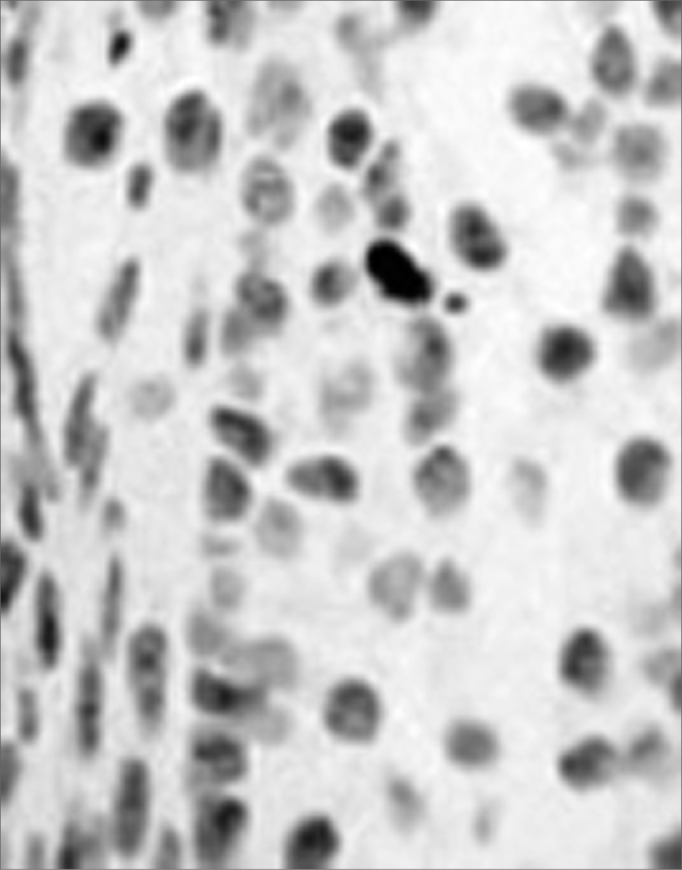
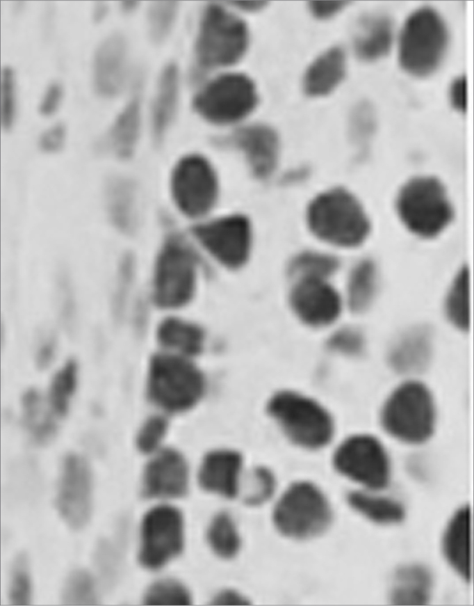
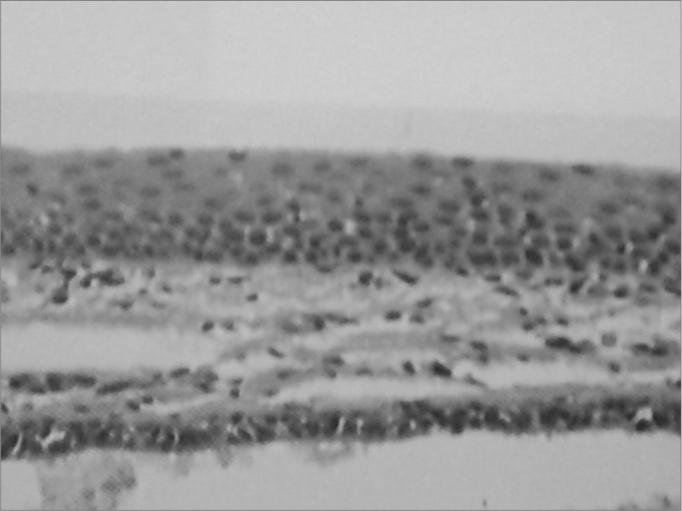
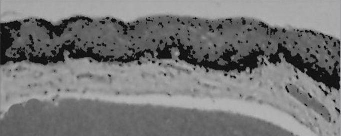
Results: We found progesterone receptors in 18 of the 19 patients. The progesterone receptors are located both in the nucleus and the cytoplasm of cells, and mainly in the basal layer. There was no report of estrogen receptors present in the vocal folds.
Conclusion: Vocal fold epithelium bears progesterone receptors, in the cytoplasm and in the nucleus. We did not find estrogen receptors in the epithelia of the vocal folds investigated.
Figures
References
-
- Schmidt BMW, Gerdes D, Feuring M. Rapid, nongenomic steroid actions: a new age? Front Neuroendocrinol. 2000;21:57–94. - PubMed
-
- Gruber CJ, Tschugguel W, Schneeberger C, Huber JC. Mechanisms of Disease: Production and Actions of Estrogens. New England J Med. 2002;346(5):340–352. - PubMed
-
- Thompson AR. Pharmacological agents with effects on voice. Am J Otolaryngol. 1995;16(1):12–18. - PubMed
-
- Woisard V, Percondani J, Serrano E, Pessey JJ. La voix de l’enfant, évolution morphologique du larynx et sés conséquences acoustiques. Laryngol Otol Rhinol. 1996;117(4):313–317. - PubMed
-
- Higgins MB, Saxman JH. Variations in vocal frequency perturbation across the menstrual cycle. J Voice. 1989;3:233–243.
Publication types
MeSH terms
Substances
LinkOut - more resources
Full Text Sources
