. 2008 Oct 3;135(1):61-73.
doi: 10.1016/j.cell.2008.07.043.

Hypothalamic IKKbeta/NF-kappaB and ER stress link overnutrition to energy imbalance and obesity

Affiliations

Hypothalamic IKKbeta/NF-kappaB and ER stress link overnutrition to energy imbalance and obesity

Xiaoqing Zhang et al. Cell. .

Abstract

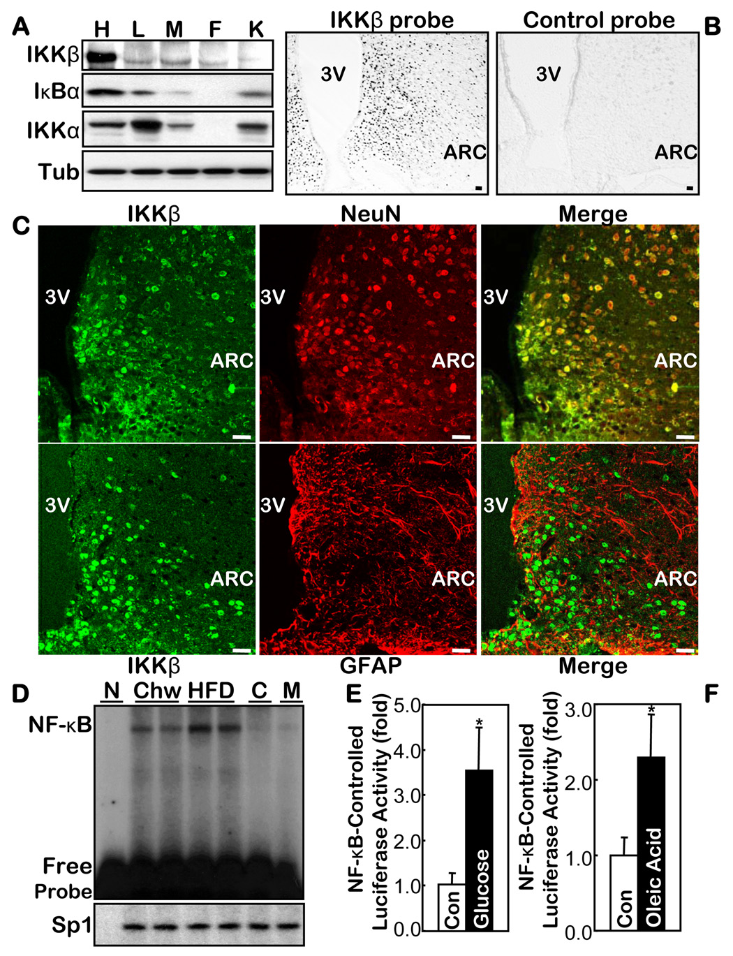
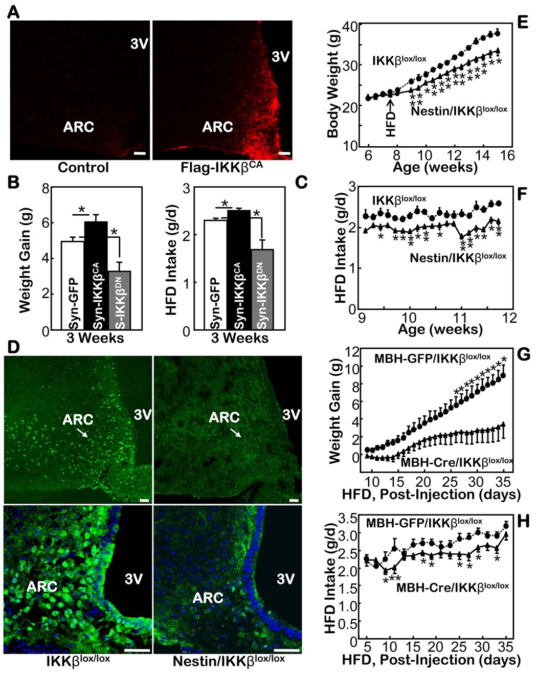
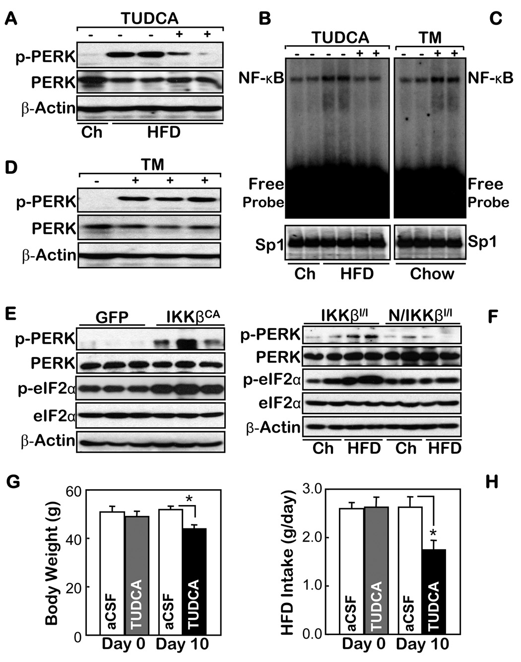
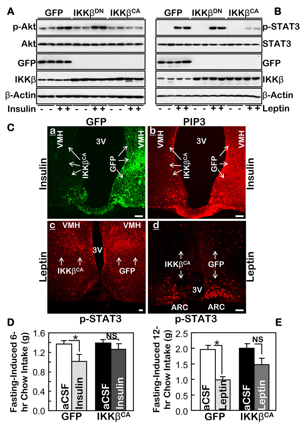
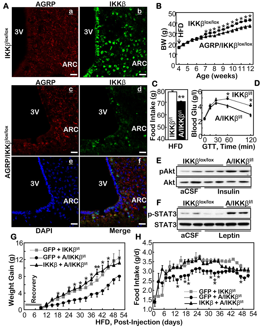
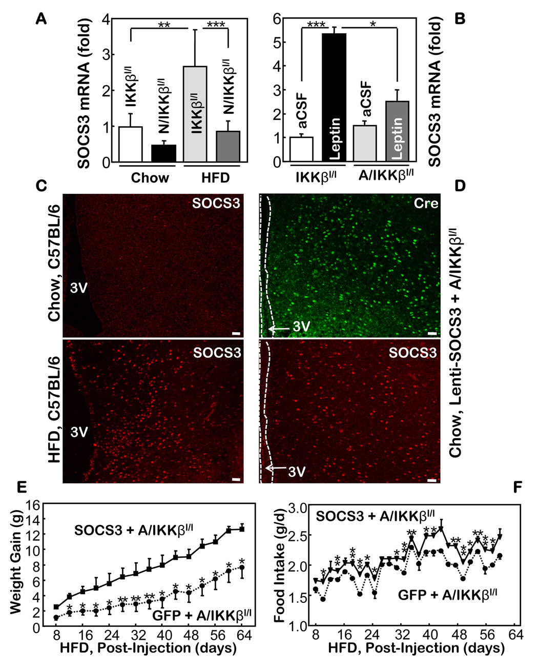
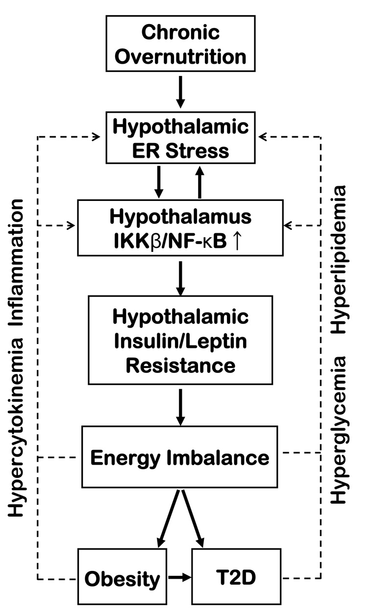
Overnutrition is associated with chronic inflammation in metabolic tissues. Whether metabolic inflammation compromises the neural regulatory systems and therefore promotes overnutrition-associated diseases remains unexplored. Here we show that a mediator of metabolic inflammation, IKKbeta/NF-kappaB, normally remains inactive although enriched in hypothalamic neurons. Overnutrition atypically activates hypothalamic IKKbeta/NF-kappaB at least in part through elevated endoplasmic reticulum stress in the hypothalamus. While forced activation of hypothalamic IKKbeta/NF-kappaB interrupts central insulin/leptin signaling and actions, site- or cell-specific suppression of IKKbeta either broadly across the brain or locally within the mediobasal hypothalamus, or specifically in hypothalamic AGRP neurons significantly protects against obesity and glucose intolerance. The molecular mechanisms involved include regulation by IKKbeta/NF-kappaB of SOCS3, a core inhibitor of insulin and leptin signaling. Our results show that the hypothalamic IKKbeta/NF-kappaB program is a general neural mechanism for energy imbalance underlying obesity and suggest that suppressing hypothalamic IKKbeta/NF-kappaB may represent a strategy to combat obesity and related diseases.


Figures

Fig. 1
Fig. 1
IKKβ/NF-κB in the hypothalamus and its relationship with overnutrition. A. Protein levels of IKKβ, IKKα and IκBα in the hypothalamus (H) vs. the peripheral organs including liver (L), skeletal muscle (M), fat (F), and kidney (K) from normal chow-fed C57BL/6 mice. Tub: tubulin. B & C. Distribution of IKKβ mRNA and protein in the hypothalamus was respectively profiled by probing the brain sections with anti-sense IKKβ oligonucleotides (B) and by co-immunostaining of IKKβ with NeuN or GFAP (C). The labeled sense sequence of IKKβ oligonucleotides was used as the control probe (B, right). Bar = 50 µm. D. DNA binding activity of NF-κB oligonucleotide and an irrelevant control (Sp1) was measured using EMSA in the hypothalamus from HFD- vs. chow-fed C57BL/6 mice. Cold competition (C) with 200-fold excess of unlabelled NF-κB oligo and mutant NF-κB probe (M) were used to assess the specificity of the NF-κB EMSA blots. N: non-specific protein (BSA). E & F. NF-κB-induced luciferase activity was measured in the hypothalamus harvested from the NF-κB reporter mice that received intra-third ventricle infusion of 1 mg glucose (E) or 40 mg oleic acid (F) over 6 hours. The controls (Con) represent the infusion of the empty vehicle or the same concentrations of sorbitol. (n = 4–5 per group, *p<0.05). 3V: third ventricle.
Fig. 2
Fig. 2
Mouse phenotypes with brain or hypothalamic IKKβ manipulations. A. Immunostaining using anti-Flag antibody for the hypothalamic sections from mice that received intra-MBH injections of Flag-IKKβCA lentivirus and the control lentivirus. Bar = 50 µm. B & C. HFD-fed C57BL/6 mice received intra-MBH injections of a lentivirus in which the synapsin (Syn, S) promoter was employed to direct the neuronal expression of IKKβCA, IKKβDN, and control GFP. Weight gain (B) and food intake (C) were followed after the mice recovered from injections and surgeries. (n = 4–6 per group, *p<0.05). D. Immunostaining of IKKβ in the hypothalamus of Nestin/IKKβlox/lox mice vs. the control IKKβlox/lox mice. Lower panels: the IKKβ staining (green) was merged with the nuclear staining of the MBH cells (blue) by DAPI. Bar = 50 µm. E & F. Body weight (E) and daily food intake (F) were measured for HFD-fed Nestin-IKKβlox/lox mice vs. littermate IKKβlox/lox mice. (n = 5–6 per group, *p<0.05). G & H. Body weight gain (G) and HFD intake (H) were measured in IKKβlox/lox mice that received MBH injections of Cre- or GFP-adenovirus (n = 11–13 per group, *p<0.05).
Figure 3
Figure 3
The association of ER stress with IKKβ/NF-κB in the hypothalamus. A–D. Normal chow (Ch)-fed mice (A–D) and HFD-fed matched mice (A & B) received intra-third ventricle infusions of (+) 25 µg TUDCA (B) or 3 µg TM (C) vs. the vehicle control (−) over 2 hours (A–D). The markers of ER stress (A & D) and the DNA binding activities of NF-κB and an irrelevant control (Sp1) (B & C) in the hypothalamus of these mice were measured using Western blot and EMSA, respectively. E & F. The ER stress markers were measured in the hypothalamus of normal chow-fed mice that received intra-MBH injections of IKKβCA- vs. GFP-lentivirus (E) and normal chow- vs. HFD-fed Nestin/IKKβlox/lox mice (N/IKKβl/l) and their controls (IKKβlox/lox mice, IKKβl/l) (F). G & H. HFD-fed mice received daily intra-third ventricle injections of TUDCA ((5 µg/d) or the empty vehicle (aCSF) for 10 days. The body weight (G) and average food intake (H) of these mice were measured before (Day 0) and upon the completion of the 10-day therapy. (n = 5 per group; *p<0.05).
Figure 4
Figure 4
IKKβ/NF-κB in the MBH mediates central insulin and leptin resistance. A & B. An adenovirus was injected to deliver IKKβCA, IKKβDN, or GFP to both MBH sides of normal C57BL/6 mice. Following surgical recovery, 24-hour-fasted mice received third-ventricle injections of either insulin (A) or leptin (B) (+), or the empty vehicle (−) (A & B). Proteins in the dissected hypothalamus were immunoblotted with the indicated antibodies. C. Adult C57BL/6 mice were injected with adenoviruses to deliver IKKβCA into one side of the MBH and GFP into the other side (a) of the same mice. Recovered mice were fasted 48 hours and then stimulated with insulin (a–b) or leptin (c–d) for 20 min via a third-ventricle cannula. Brain sections across the MBH were directly examined for GFP (a), and immunostained with the indicated primary antibodies (b–d). Arrows indicate the regions in which the injected genes (IKKβCA and GFP) were expressed. Bar = 100 µm. D & E. An adenovirus was injected into adult C57BL/6 mice to deliver either IKKβCA to both sides of the MBH of one group of mice or GFP to both sides of the MBH of another group of mice. Recovered mice fasted for 4 hours received third-ventricle injections of insulin (D), leptin (E), or the vehicle (aCSF) (D & E). Food intake during the indicated time periods following the insulin or leptin injections was measured. (Insulin: n = 12 per group; Leptin: n = 7–8 per group; *p<0.05). NS = non significant.
Figure 5
Figure 5
Anti-obesity effects of knocking out IKKβ in the hypothalamic AGRP neurons. A. AGRP (a & c) and IKKβ (b & d) in the hypothalamus of the control mice (IKKβlox/lox mice) (a & b) vs. AGRP-IKKβlox/lox mice (c & d) were immunostained. Immunostaining of AGRP (c) and IKKβ (d) and the nuclear staining by DAPI (e) in the hypothalamus of AGRP-IKKβlox/lox mice were merged to display the absence of IKKβ in AGRP neurons (f). Bar = 25 µm. B–D. Body weight (BW) (B), HFD intake (C), and glucose tolerance (GTT) (D) were tested in HFD-fed AGRP-IKKβlox/lox mice and IKKβlox/lox mice. (n = 12–14 per group, *p<0.05). Glu, Glucose. E & F. HFD-fed AGRP-IKKβlox/lox mice and IKKβlox/lox mice were fasted and received third-ventricle injections of insulin (5 mU) (E), leptin (10 µg) (F), or the vehicle (aCSF) (E & F). Insulin-induced Akt phosphorylation (pAkt) (E) and leptin-induced STAT3 phosphorylation (pSTAT3) (F) were determined using Western blots. G & H. Adenovirus was injected to deliver IKKβ to the both MBH sides of AGRP-IKKβlox/lox mice. For the controls, adenovirus was injected to deliver GFP to the both MBH sides of AGRP-IKKβlox/lox mice and to the both MBH sides of IKKβlox/lox mice. Body weight (G) and food intake (H) in these mice were followed for 8 weeks following the viral injections (n = 5 per group; **p<0.01). C–H. IKKβl/l: IKKβlox/lox mice; A/IKKβl/l: AGRP-IKKβlox/lox mice.
Figure 6
Figure 6
The role of SOCS3 for the anti-obesity phenotype of AGRP-IKKβlox/lox mice. A. Relative mRNA levels of SOCS3 in the hypothalamus of HFD- and chow-fed Nestin-IKKβlox/lox mice and IKKβlox/lox mice. B. Relative mRNA levels of SOCS3 in the hypothalamus of chow-fed IKKβlox/lox mice and AGRP-IKKβlox/lox mice that received third-ventricle injections of leptin. (n = 4–6 per group; *p<0.05, **p<10−2, ***p<10−3). C. SOCS3 immunostaining in the MBH of chow- vs. HFD-fed C57BL/6 mice. D. Immunostaining of Cre (green) and SOCS3 (red) in chow-fed AGRP-IKKβlox/lox mice following intra-MBH injections of SOCS3 lentivirus. E & F. SOCS3 lentivirus (SOCS3) or GFP lentivirus (GFP) was injected into the MBH of HFD-fed AGRP-IKKβlox/lox mice; body weight (E) and HFD intake (F) of injected mice were recorded. (n = 4–8 per group; *p<0.05, **p<0.01). Bar = 50 µm. A–F. IKKβl/l: IKKβlox/lox mice; N/IKKβl/l: Nestin-IKKβlox/lox mice; A/IKKβl/l: AGRP-IKKβlox/lox mice.
Figure 7
Figure 7
Schematic of the proposed role of hypothalamic IKKβ/NF-κB and ER stress in obesity-related disease pathways. We propose that IKKβ/NF-κB connects with ER stress in the hypothalamus and translates overnutrition into central insulin and leptin resistance, leading to the development of obesity and T2D (solid arrows). We also further postulate that, as obesity and T2D progress, IKKβ/NF-κB participates in cumulative feedback loops (broken arrows).

Comment in

References

    1. Arkan VC, Hevener AL, Greten FR, Maeda S, Li ZW, Long JM, Wynshaw-Boris A, Poli G, Olefsky J, Karin M. Nat. Med. 2005;11:191–198. - PubMed
    1. Balthasar N, Coppari R, McMinn J, Liu SM, Lee CE, Tang V, Kenny CD, McGovern RA, Chua SC, Jr, Elmquist JK, Lowell BB. Leptin receptor signaling in POMC neurons is required for normal body weight homeostasis. Neuron. 2004;42:983–991. - PubMed
    1. Bates SH, Stearns WH, Dundon TA, Schubert M, Tso AW, Wang Y, Banks AS, Lavery HJ, Haq AK, Maratos-Flier E, Neel BG, Schwartz MW, Myers MG., Jr STAT3 signalling is required for leptin regulation of energy balance but not reproduction. Nature. 2003;421:856–859. - PubMed
    1. Bence KK, Delibegovic M, Xue B, Gorgun CZ, Hotamisligil GS, Neel BG, Kahn BB. Neuronal PTP1B regulates body weight, adiposity and leptin action. Nat. Med. 2006;12:917–924. - PubMed
    1. Bruning JC, Gautam D, Burks DJ, Gillette J, Schubert M, Orban PC, Klein R, Krone W, Muller-Wieland D, Kahn CR. Role of brain insulin receptor in control of body weight and reproduction. Science. 2000;289:2122–2125. - PubMed

Publication types

MeSH terms