

Segregated hemispheric pathways through the optic chiasm distinguish primates from rodents
- PMID: 18854206
- PMCID: PMC2736912
- DOI: 10.1016/j.neuroscience.2008.09.021
Segregated hemispheric pathways through the optic chiasm distinguish primates from rodents
Abstract
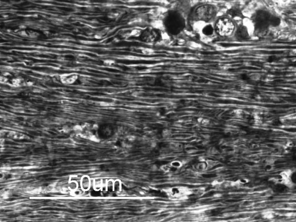
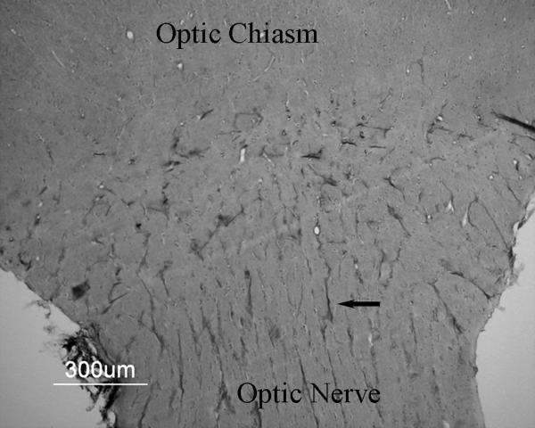
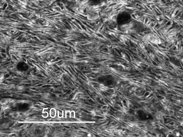
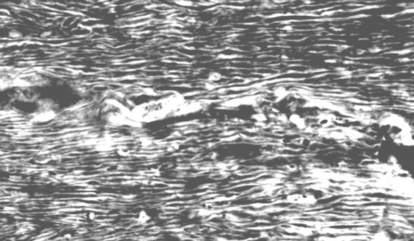
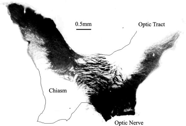
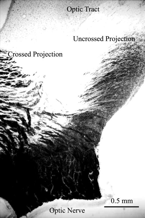
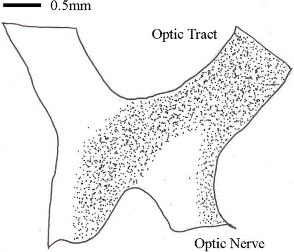
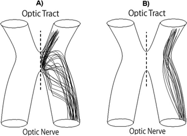
At the optic chiasm retinal fibers either cross the midline, or remain uncrossed. Here we trace hemispheric pathways through the marmoset chiasm and show that fibers from the lateral optic nerve pass directly toward the ipsilateral optic tract without any significant change in fiber order and without approaching the midline, while those from medial regions of the nerve decussate directly. Anterograde labeling from one eye shows that the two hemispheric pathways remain segregated through the proximal nerve and chiasm with the uncrossed confined laterally. Retrograde labeling from the optic tract confirms this. This clearly demonstrates that hemispheric pathways are segregated through the primate chiasm. Previous chiasmatic studies have been undertaken mainly on rodents and ferrets. In these species there is a major change in fiber order pre-chiasmatically, where crossed and uncrossed fibers mix, reflecting their embryological history when all fibers approach the midline prior to their commitment to innervate either hemisphere. This pattern was thought to be common to placental mammals. In marsupials there is no change in fiber order and uncrossed fibers remain confined laterally through nerve and chiasm, again, reflecting their developmental history when all uncrossed fibers avoid the midline. Recently it has been shown that this distinction is not a true dichotomy between placental mammals and marsupials, as fiber order in tree shrews and humans mirrors the marsupial pattern. Architectural differences in the mature chiasm probably reflect different developmental mechanisms regulating pathway choice. Our results therefore suggest that both the organization and development of the primate optic chiasm differ markedly from that revealed in rodents and carnivores.
Figures
Similar articles
-
Chiasm formation in man is fundamentally different from that in the mouse.Eye (Lond). 2007 Oct;21(10):1264-70. doi: 10.1038/sj.eye.6702839. Eye (Lond). 2007. PMID: 17914429 Review.
-
Early midline interactions are important in mouse optic chiasm formation but are not critical in man: a significant distinction between man and mouse.Eur J Neurosci. 2006 Jun;23(11):3034-42. doi: 10.1111/j.1460-9568.2006.04827.x. Eur J Neurosci. 2006. PMID: 16819992
-
First evidence of diversity in eutherian chiasmatic architecture: tree shrews, like marsupials, have spatially segregated crossed and uncrossed chiasmatic pathways.J Comp Neurol. 1998 Jan 12;390(2):183-93. doi: 10.1002/(sici)1096-9861(19980112)390:2<183::aid-cne2>3.0.co;2-y. J Comp Neurol. 1998. PMID: 9453663
-
Distinctive pattern of organisation in the retinofugal pathway of a marsupial: II. Optic chiasm.J Comp Neurol. 1992 Nov 1;325(1):57-67. doi: 10.1002/cne.903250106. J Comp Neurol. 1992. PMID: 1484119
-
Architecture of the optic chiasm and the mechanisms that sculpt its development.Physiol Rev. 2001 Oct;81(4):1393-414. doi: 10.1152/physrev.2001.81.4.1393. Physiol Rev. 2001. PMID: 11581492 Review.
Cited by
-
Development of the retina and optic pathway.Vision Res. 2011 Apr 13;51(7):613-32. doi: 10.1016/j.visres.2010.07.010. Epub 2010 Jul 18. Vision Res. 2011. PMID: 20647017 Free PMC article. Review.
-
Altered anterior visual system development following early monocular enucleation.Neuroimage Clin. 2013 Nov 1;4:72-81. doi: 10.1016/j.nicl.2013.10.014. eCollection 2014. Neuroimage Clin. 2013. PMID: 24319655 Free PMC article.
-
Human Pluripotent Stem Cell-Derived Retinal Ganglion Cells: Applications for the Study and Treatment of Optic Neuropathies.Curr Ophthalmol Rep. 2015 Sep;3(3):200-206. doi: 10.1007/s40135-015-0081-9. Epub 2015 Aug 7. Curr Ophthalmol Rep. 2015. PMID: 26618076 Free PMC article. No abstract available.
-
Vitrectomy and ILM peeling in rhesus macaque: pitfalls and tips for success.Eye (Lond). 2023 Aug;37(11):2257-2264. doi: 10.1038/s41433-022-02327-5. Epub 2022 Nov 28. Eye (Lond). 2023. PMID: 36443497 Free PMC article.
References
-
- Baker GE. Prechiasmatic reordering of fibre diameter classes in the retinofugal pathway of the ferret. Eur J Neurosci. 1990;2:24–33. - PubMed
-
- Baker GE, Jeffery G. Distribution of uncrossed axons along the course of the optic nerve and chiasm of rodents. J Comp Neurol. 1989;289:455–461. - PubMed
-
- Baker GE, Reese BE. Chiasmatic course of temporal retinal axons in the developing ferret. J Comp Neurol. 1993;330:95–104. - PubMed
-
- Bancroft JD, Stevens A, editors. Theory and Practice of Histological Techniques. Churchill Livingstone; Edinburgh: 1990.
-
- Day AL. Aneurysms of the ophthalmic segment: a clinical and anatomical analysis. J Neurosurg. 1990;72:677–691. - PubMed
Publication types
MeSH terms
Substances
Grants and funding
LinkOut - more resources
Full Text Sources
