. 2008 Oct 16;4(4):325-36.
doi: 10.1016/j.chom.2008.08.001.

An inhibitor of gram-negative bacterial virulence protein secretion

Affiliations

An inhibitor of gram-negative bacterial virulence protein secretion

Heather B Felise et al. Cell Host Microbe. .

Abstract

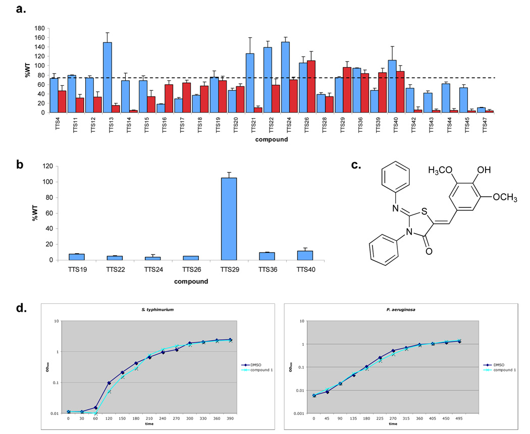
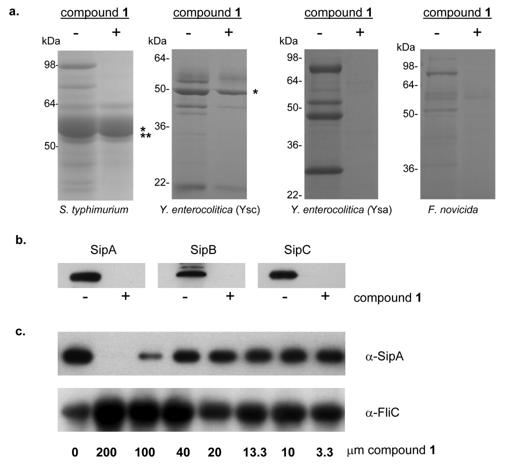
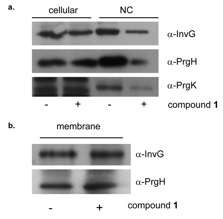
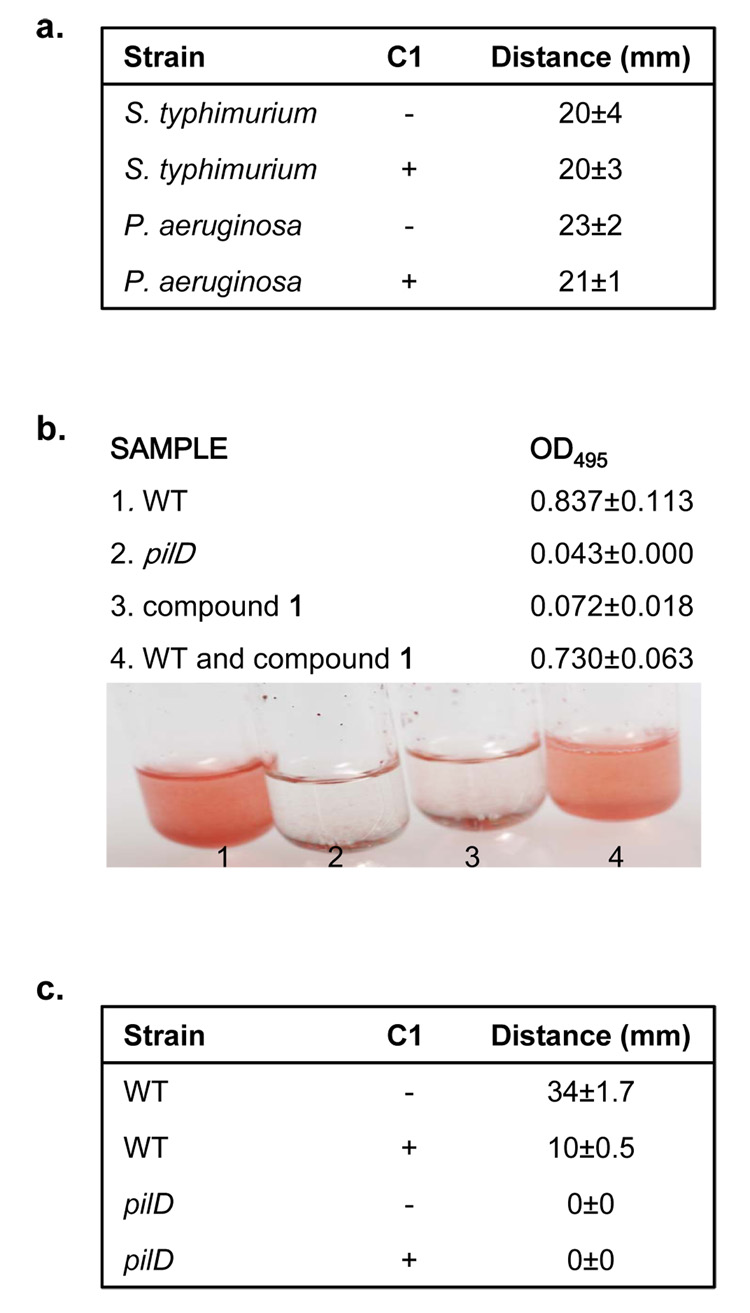
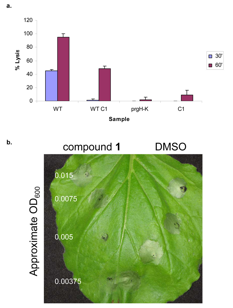
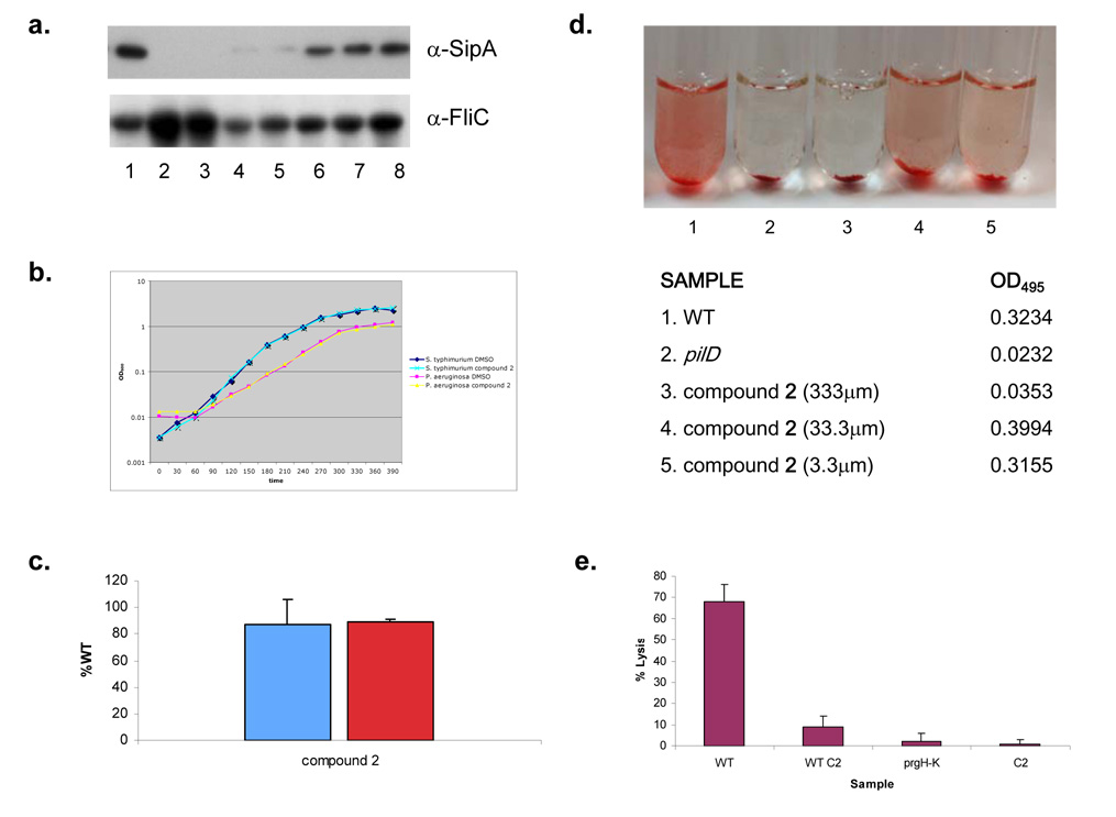
Bacterial virulence mechanisms are attractive targets for antibiotic development because they are required for the pathogenesis of numerous global infectious disease agents. The bacterial secretion systems used to assemble the surface structures that promote adherence and deliver protein virulence effectors to host cells could comprise one such therapeutic target. In this study, we developed and performed a high-throughput screen of small molecule libraries and identified one compound, a 2-imino-5-arylidene thiazolidinone that blocked secretion and virulence functions of a wide array of animal and plant Gram-negative bacterial pathogens. This compound inhibited type III secretion-dependent functions, with the exception of flagellar motility, and type II secretion-dependent functions, suggesting that its target could be an outer membrane component conserved between these two secretion systems. This work provides a proof of concept that compounds with a broad spectrum of activity against Gram-negative bacterial secretion systems could be developed to prevent and treat bacterial diseases.


Figures

Figure 1
Figure 1. Identification of an inhibitor of T3S in S. typhimurium
a,β-galactosidase activity from an flhC-lacZ transcriptional fusion in S. typhimurium when grown in the presence of small molecules (~380 µm) identified in the HTS screen. Activity was recorded as a percent of WT (blue bars), i.e. the amount of β-galactosidase activity observed when bacteria are grown in the absence of compound. Three independent experiments were performed and the mean standard deviations are shown. Alkaline phosphotase activity of a PrgH-PhoA translational fusion in S. typhimurium when grown in the presence of small molecules (~380 m) identified in the HTS screen (red bars). The dashed line (---) marks 60% of WT activity, the level used to choose compounds for further study. b, β-galactosidase activity from an iagB-lacZ transcriptional fusion in S. typhimurium when grown in the presence of small molecules (380 µm) with levels of flhC-lacZ and PrgH-PhoA expression greater than 60%. As above, activity was recorded as a percent of WT. Three independent experiments were performed and the mean standard deviations are shown. c, Chemical structure of TTS29, renamed Compound 1. d, Growth curves for S. typhimurium and P. aeruginosa in LB medium with compound 1 or an equal volume of the solvent.
Figure 2
Figure 2. Phenotypic effects of compound 1 on type III secretion
a, Bacterial cultures were grown in the absence (−) or presence (+) of 380 µM of compound 1. Secreted proteins were TCA precipitated and separated by 12.5% SDS-PAGE and stained with Coomaisse Blue. The flagellin proteins from S. typhimurium and Y. enterocolitica are marked (*) as well as the associated flagellar cap protein in S. typhimurium (**). b, Western Blots of the secreted proteins, SipA, B and C from S. typhimurium. c, Western Blots of secreted SipA protein as well as the flagellar filament protein, FliC in supernatants from S. typhimurium grown in LB alone or with decreasing concentrations of compound 1.
Figure 3
Figure 3. Phenotypic effects of compound 1 on type III secretion needle complex assembly
a, Type III secretion needle complexes (NC) were purified from S. typhimurium grown in the absence (−) or presence (+) of 380 µM of compound 1. Proteins were separated by SDS-PAGE and Western Blotted with antibodies to InvG, PrgH and PrgK. b, Total membrane fractions were isolated from S. typhimurium grown in the absence (−) or presence (+) of 380 µM of compound 1. Proteins were separated by SDS-PAGE and Western Blotted with antibodies to InvG and PrgH.
Figure 4
Figure 4. Phenotypic effects of compound 1 on alternate secretion systems
a, Motility assays of S. typhimurium and P. aeruginosa in the absence (−) or presence (+) of 380 µM of compound 1. Bacteria were stabbed into motility plates and incubated at 37°C for 6 hours. The diameter of the ring was measured for three independent replicates and the mean standard deviations are shown. b, Elastolytic activity for 18 hour culture supernatants of P. aeruginosa grown in the absence (WT; 1) or presence of 380 µM of compound 1 (compound 1; 3) was determined using elastin Congo Red as a substrate. Elastase activity was measured as a change in OD495 and can be observed as an increase in the red color of the sample. As a negative control elastase activity was determined for the culture supernatant of a P. aeruginosa T2S mutant (pilD; 2). As a control for inhibition of elastase activity by the compound, compound 1 was added directly to culture supernatants from P. aeruginosa grown in the absence of compound and assayed for elastolytic activity (WT and compound 1; 4). Pictures and the corresponding OD495 of samples are shown. c, Twitching assays of P. aeruginosa in either the absence (−) or presence (+) of 380 µM of compound 1. Bacteria were stabbed into LB plates with 1.0% agar and incubated for 2 days at room temperature. Growth media was removed and the bacteria were stained with crystal violet for better visualization.
Figure 5
Figure 5. Inhibition of bacterial virulence phenotypes by compound 1
a, Mouse bone marrow macrophages (BMM) from Balb/c mice were infected with WT S. typhimurium grown in the absence (WT) or presence of 380 µM of compound 1 (WT C1) to SPI1 inducing conditions. A T3S genetic mutant (prgH–K) was used as a negative control. To confirm that compound 1 was not itself cytotoxic to the macrophages, compound without bacteria (C1) was added at a concentration equal to the experimental samples. Bacteria were added at an MOI of 40 and cytotoxicity was assessed by LDH release at 30 minutes and 60 minutes after infection. Assays were performed in quadruplicate and mean standard deviations are shown. b, P. syringae and compound 1 were co-inoculated on non-host tobacco plants and monitored for HR. Varying concentrations of bacteria were added, while the amount of compound or solvent (v/v) remained constant. Four independent experiments were performed and a representative experiment is shown. Tissue collapse near the point of inoculation for either compound 1 or solvent alone is due to tissue damage caused during injection of compounds by a blunt-ended syringe.
Figure 6
Figure 6. Characterization of secretion inhibition by compound 2
a, Western Blots of secreted SipA protein as well as the flagellar filament protein, FliC in supernatants from S. typhimurium grown in LB alone or with decreasing concentrations of compound 2. b, Growth curves for S. typhimurium and P. aeruginosa in LB medium with compound 2 or an equal volume of solvent. c, β-galactosidase activity (blue bar) from an iagB-lacZ transcriptional fusion in S. typhimurium or the amount of alkaline phosphotase activity (red bar) observed from a PrgH-PhoA translational fusion in S. typhimurium when grown in the presence of compound 2 (100 µm). Enzymatic activity was recorded as a percent of WT, i.e. the amount of activity observed in the absence of compound. Three independent experiments were performed and the mean standard deviations are shown. d, Elastolytic activity for 18 hour culture supernatants of P. aeruginosa grown in the absence (1) or presence of compound 2 at varying concentrations (3, 4 & 5) was determined using elastin Congo Red as a substrate. Elastase activity was measured as a change in OD495 and can be observed as an increase in the red color of the sample. As a negative control elastase activity was determined for the culture supernatant of a P. aeruginosa T2S mutant (pilD; 2). Pictures and the corresponding OD495 of samples are shown. e, Mouse BMMs were infected with WT S. typhimurium grown in the absence (WT) or presence of 100 µm of compound 2 (WT C2), a T3S mutant (prgH–K) or compound 2 without bacteria (C2). Bacteria were added at an MOI of 40 and cytotoxicity was assessed by LDH release at 60 minutes. Assays were performed in quadruplicate and mean standard deviations are shown.

References

    1. Bitter W. Secretins of Pseudomonas aeruginosa: large holes in the outer membrane. Arch Microbiol. 2003;179:307–314. - PubMed
    1. Cianciotto NP. Type II secretion: a protein secretion system for all seasons. Trends Microbiol. 2005;13:581–588. - PubMed
    1. Cornelis GR. The type III secretion injectisome. Nat Rev Microbiol. 2006;4:811–825. - PubMed
    1. Cornelis GR, Van Gijsegem F. Assembly and function of type III secretory systems. Annu Rev Microbiol. 2000;54:735–774. - PubMed
    1. Datsenko KA, Wanner BL. One-step inactivation of chromosomal genes in Escherichia coli K-12 using PCR products. Proc Natl Acad Sci. 2000;97:6640–6645. - PMC - PubMed

Publication types

MeSH terms