. 2008 Oct 16;4(4):387-97.
doi: 10.1016/j.chom.2008.09.001.

Mechanism of induction and suppression of antiviral immunity directed by virus-derived small RNAs in Drosophila

Affiliations

Mechanism of induction and suppression of antiviral immunity directed by virus-derived small RNAs in Drosophila

Roghiyh Aliyari et al. Cell Host Microbe. .

Abstract

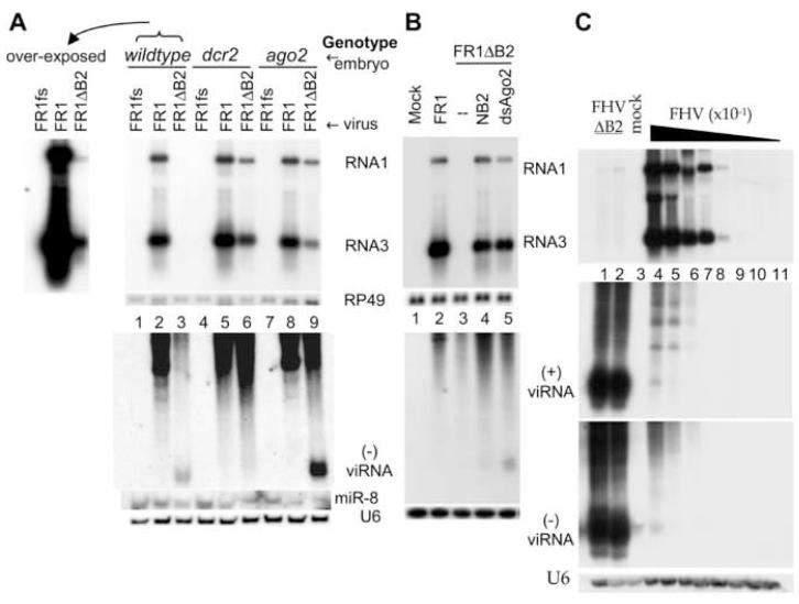
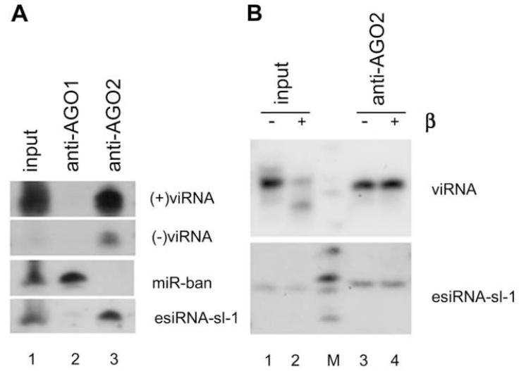
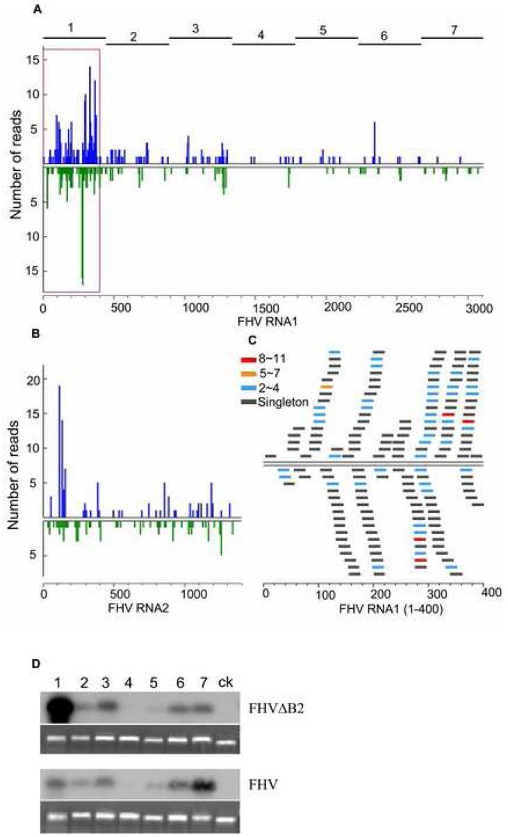
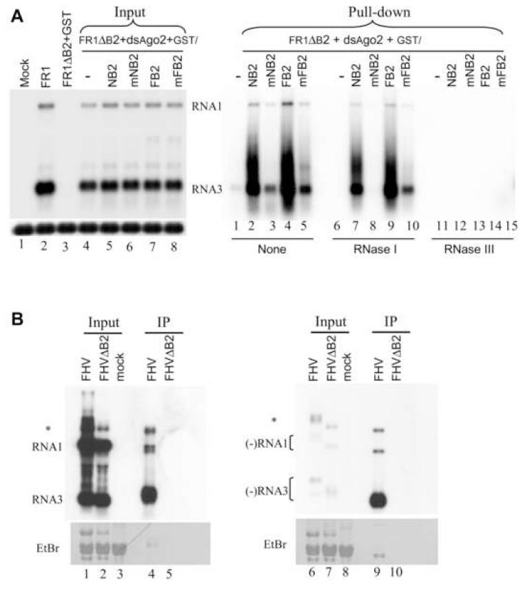
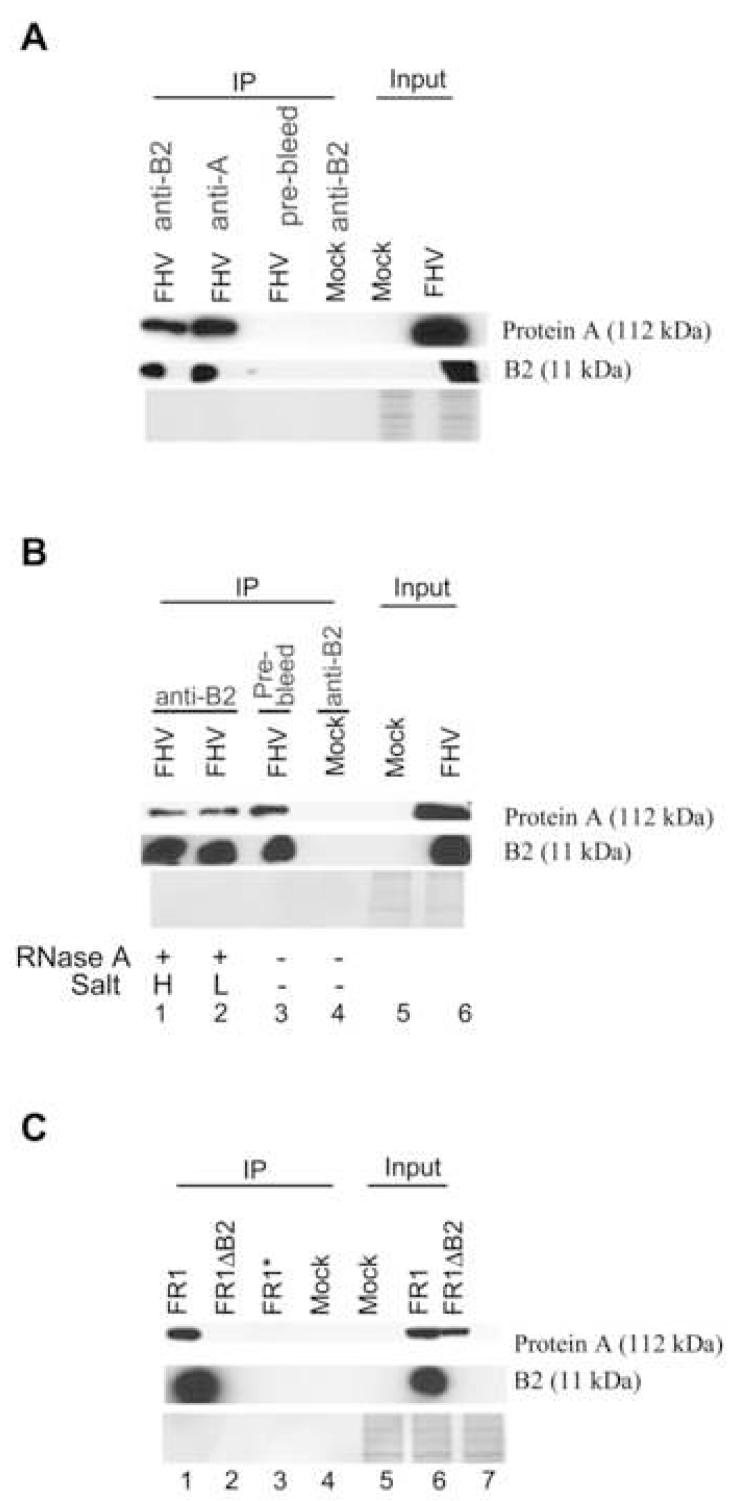
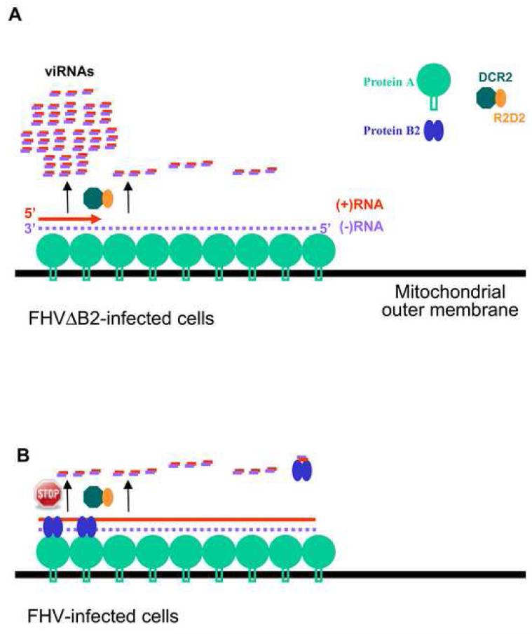
The small RNA-directed viral immunity pathway in plants and invertebrates begins with the production by Dicer nuclease of virus-derived siRNAs (viRNAs), which guide specific antiviral silencing by Argonaute protein in an RNA-induced silencing complex (RISC). Molecular identity of the viral RNA precursor of viRNAs remains a matter of debate. Using Flock house virus (FHV) infection of Drosophila as a model, we show that replication of FHV positive-strand RNA genome produces an approximately 400 bp dsRNA from its 5' terminus that serves as the major Dicer-2 substrate. ViRNAs thus generated are loaded in Argonaute-2 and methylated at their 3' ends. Notably, FHV-encoded RNAi suppressor B2 protein interacts with both viral dsRNA and RNA replicase and inhibits production of the 5'-terminal viRNAs. Our findings, therefore, provide a model in which small RNA-directed viral immunity is induced during the initiation of viral progeny (+)RNA synthesis and suppressed by B2 inside the viral RNA replication complex.


Figures

Figure 1
Figure 1
DCR2-dependent production of viRNA is inhibited by B2. (A, B) Accumulation of viral RNA1 and RNA3 (upper panel) as well as viRNAs (lower panel) 48 hours after micro-injection into embryos (A) or transcriptional induction in S2 cells (B) of FHV RNA1 (FR1), B2-deficient RNA1 (FR1ΔB2) or replication-defective FR1fs, which contains a frameshift mutation in the viral RdRP gene. Top left of panel A shows an overexposure of lanes 1-3. In panel B, pFR1ΔB2 was co-transfected with an additional plasmid encoding B2 of NoV (lane 4), or dsRNA targeting AGO2 (lane 5) or LacZ (lane 3). viRNAs were detected by eleven 40-nt oligos hybridizing to the (-)-strand of the coding region for ORF B2. (C) Accumulation of viral RNA1 and RNA3 as well as (+) and (-) viRNAs in S2 cells 72 hours after inoculation with virions of a B2-deficient FHV mutant (FHVΔB2, lanes 1-2) or FHV in a series of 10-fold dilutions (lanes 4-11). The same samples were analyzed for (+) and (-) viRNAs in two identical gels by using oligo probes targeting the (+) and (-)-strand of the 5′-terminal region of RNA1. Equal loading was monitored by probing for RP49 or U6.
Figure 2
Figure 2
Characterization of viRNAs in the infected cells. (A) viRNAs are associated with AGO2 but not AGO1 in infected Drosophila cells. AGO1 and AGO2 were immunoprecipitated respectively by specific antibodies four days after inoculation with FHVΔB2 virions and bound small RNAs were analyzed by Northern blot hybridizations using probes specific to (+) and (-) viRNAs, miR-ban and esiRNA-sl-1. (B) viRNAs loaded in AGO2 were resistant to peridate oxidation and beta elimination treatments (β) whereas viRNAs in the input before immunoprecipitation were partially sensitive. The samples used were identical to lanes 1 and 3 of (A).
Figure 3
Figure 3
Examining the population of viRNAs produced in Drosophila cells. (A, B) Profiles of viRNAs cloned by the 5′-ligation independent method from S2 cells abortively infected with FHVΔB2. Number of viRNA reads was plotted to the positive-(top) and negative- (bottom) strand of RNA1 (A) and RNA2 (B) with 5-nt windows. (C) A close-up view of the distribution and abundance of (+) and (-) viRNAs in the 5′-terminal 400-nt region of RNA1. 264 reads were from this region including 153 (+)viRNAs (58%, top) and 111 (-)viRNAs (42%, bottom). Counts of distinct viRNAs are shown by color-coded bars. (D) Relative abundance of viRNAs targeting the 7 evenly divided regions of RNA1 (shown on top of each lane and of the graph in panel A) produced in S2 cells infected with FHV (bottom panel) or FHVΔB2 (top panel). Equal amount (300 ng) of the seven FHV RNA1-specific DNA fragments and a control LacZ fragment was fractionated and hybridized to the labeled total 20- to 24-nt viRNAs gel purified from approximately 300 μg of total RNA extracted from S2 cells 4 days after inoculation with virions of FHV or FHVΔB2. Equal loading was shown by staining with ethidium bromide.
Figure 4
Figure 4
Detection of B2-dsRNA complexes in infected Drosophila cells. (A) Northern blot analysis of viral RNAs in S2 cells before (input lanes of left panel) and after (right panel) GST pulldown. S2 cells were co-transfected with pFR1ΔB2 and dsRNA of AGO2 plus a plasmid expressing GST alone (lane 1 of left panel and lanes 1, 6 and 11 of right panel), GST-tagged wildtype (lane 5 and 7 of left panel and lanes 2, 4, 7, 9, 12 and 14 of right panel) or (R→Q) mutant B2 (lane 6 and 8 of left panel and lanes 3, 5, 8, 10, 13 and 15 of right panel) of FHV and NoV. 5% of total RNA before GST pulldown was analyzed in the left panel. Right panel shows the RNA pulled down by GST with treatment of RNase I (lanes 6-10), RNase III (lanes 11-15) or without RNase treatment (lanes 1-5). (B) Northern blot analysis of viral RNAs in mock-infected S2 cells and S2 cells infected with virions of FHV or FHVΔB2 before (input) and after coimmunoprecipitation (IP) with the antibody to B2 of FHV. S2 cells were pre-treated with dsRNA of AGO2 before inoculation with FHVΔB2 virions, which is essential to ensure successful infection. The strand-specific probes recognized (+) and (-)-strand of FHV RNA1 and RNA3, respectively, each contained eleven 5′-labeled, 40-nt single-strand DNA oligos hybridizing to the (+) and (-)-strand of the ORF B2 coding region. The RNA species that migrated at the positions of FHV RNA1 and RNA3 were marked. Asterisk (*) indicates an RNA species that may correspond to the homodimer of RNA1 implicated in FHV replication. Methylene blue staining of the filter was shown at the bottom.
Figure 5
Figure 5
Specific interaction of B2 and viral RdRP in vivo. (A, B) Coimmunoprecipitation of B2 and protein A of FHV in FHV-infected cells. Total crude protein extracts (Input) prepared 12 hours after mock inoculation or inoculation with FHV virions were immunoprecipitated (IP) with polyclonal antibody to B2 or protein A. A pre-immune antibody was used as a negative control. Western blots were analyzed with the same antibodies to detect coimmunoprecipitated proteins. The proteins coimmunoprecipitated by the B2 antibody were treated with RNase A under high (H) or low salt concentrations (L) before fractionation and Western blot analysis (lanes 1-2 of panel B). (C) Interaction of B2 and protein A in S2 cells in which RNA1 self-replicates in absence of RNA2. Total crude protein extracts (Input) prepared in S2 cells 48 hours after induction of viral RNA replication were immunoprecipitated (IP) with either polyclonal antibody to B2 (lanes 1 and 2) or the pre-immune antibody (lane 3). Mocktransfected S2 cells were used as a negative control (lane 4). As described for Figure 1B, AGO2 was depleted by dsRNA in S2 cells transfected with pFR1ΔB2 to ensure robust replication of FR1ΔB2 (lane 2).
Figure 6
Figure 6
Model for the induction (A) and suppression (B) of the Drosophila RNAi immunity by FHV. Asymmetric RNA synthesis in the replication of (+)RNA viruses involves multiple initiation of the progeny (+)RNA synthesis on the low abundant (-)RNA template complexed with the viral RdRP and other host factors. The resulting dsRNA of approximate 400-nt in length formed between the 5′-terminal nascent progeny (+)RNA1 and the (-)RNA1 template in FHV-infected cells, termed the initiating vRI-dsRNA, serves as substrates of DCR2. This results in the predominant production of 5′-terminal viRNAs, thereby triggering the RNAi-mediated viral immunity and abortive infection by FHVΔB2. In addition to binding to viRNAs, B2 is part of the viral RNA replication complex by direct interactions with viral RdRP (protein A) and vRI-dsRNA and inhibits DCR2-dependent production of viRNAs, thus ensuring successful infection by FHV. We propose that sequestering the initiating vRI-dsRNA and inhibiting their processing into the 5′-terminal viRNAs by B2 play a particularly important role in the suppression of the viral immunity.

References

    1. Ahlquist P. Parallels among positive-strand RNA viruses, reverse-transcribing viruses and double-stranded RNA viruses. Nat Rev Microbiol. 2006;4:371–382. - PMC - PubMed
    1. Baulcombe D. RNA silencing in plants. Nature. 2004;431:356–363. - PubMed
    1. Bouche N, Lauressergues D, Gasciolli V, Vaucheret H. An antagonistic function for Arabidopsis DCL2 in development and a new function for DCL4 in generating viral siRNAs. EMBO J. 2006;25:3347–3356. - PMC - PubMed
    1. Chapman EL, Carrington JC. Specialization and evolution of endogenous small RNA pathways. Nat Rev Gene. 2007;7:844–96. - PubMed
    1. Chao JA, Lee JH, Chapados BR, Debler EW, Schneemann A, Williamson JR. Dual modes of RNA-silencing suppression by Flock House virus protein B2. Nat Struct Mol Biol. 2005;12:952–957. - PubMed

Publication types

MeSH terms