

Immunodiagnostic capabilities of anti-free immunoglobulin light chain monoclonal antibodies
- PMID: 18854262
- PMCID: PMC2620173
- DOI: 10.1309/AJCPNS6K1CYJPDBA
Immunodiagnostic capabilities of anti-free immunoglobulin light chain monoclonal antibodies
Abstract
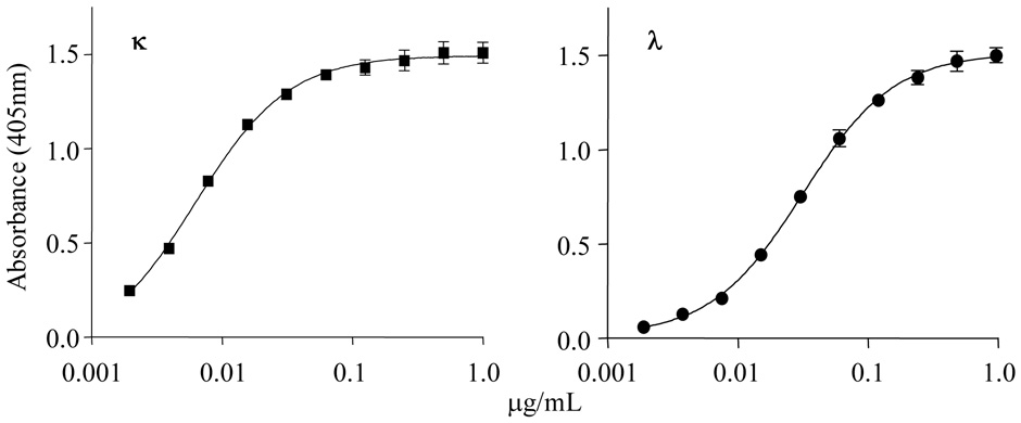
Overproduction of plasma cell-derived monoclonal free kappa or lambda immunoglobulin light chains (FLCs) is a hallmark of multiple myeloma, AL amyloidosis, and light chain deposition disease. Because these components serve as unique cellular and serologic biomarkers, their detection and quantitation has diagnostic, therapeutic, and prognostic import. In this regard, we have developed monoclonal antibodies (mAbs) that specifically recognize the kappa or lambda FLC products of all known human variable and constant region light chain genes. We now report the results of our studies that have demonstrated the capability of these reagents to measure, in a modified fluid-phase capture enzyme-linked immunosorbent assay (ELISA), serum kappa and lambda FLCs at concentrations as low as 5 and 15 ng/mL, respectively. The mAb-based ELISA has greater sensitivity and reproducibility than does the commercially available immunoturbidimetric assay that uses polyclonal anti-FLC antibodies. In addition, the mAbs can immunostain monoclonal FLC-producing plasma cells and pathologic light chain-related amyloid and nonfibrillar tissue deposits. Our anti-FLC mAbs, with their high degree of reactivity and versatility, may provide an invaluable tool in the diagnosis and management of light chain-associated disease.
Figures


Comment in
-
Monoclonal vs polyclonal free light chain assays.Am J Clin Pathol. 2009 Jun;131(6):901-2; author reply 902-3. doi: 10.1309/AJCPBWZD3EFWHNLL. Am J Clin Pathol. 2009. PMID: 19461100 No abstract available.
References
-
- Solomon A. Monoclonal immunoglobulins as biomarkers of cancer. In: Sell S, editor. Cancer Markers: Developmental and Diagnostic Significance. Clifton, NJ: Humana Press; 1980. pp. 57–87.
-
- Solomon A, Weiss DT, Herrera GA. Renal diseases associated with multiple myeloma and related plasma cell dyscrasias. In: Berenson JR, editor. Biology and Management of Multiple Myeloma. Totowa, NJ: Humana Press; 2004. pp. 281–302.
-
- Axiak SM, Krishnamoorthy L, Guinan J, Raison RL. Quantitation of free kappa light chains in serum and urine using a monoclonal antibody based inhibition enzyme-linked immunoassay. J Immunol Methods. 1987;99:141–147. - PubMed
-
- Nelson M, Brown RD, Gibson J, Joshua DE. Measurement of free kappa and lambda chains in serum and the significance of their ratio in patients with multiple myeloma. Brit J Haematol. 1992;81:223–230. - PubMed
Publication types
MeSH terms
Substances
Grants and funding
LinkOut - more resources
Full Text Sources
Other Literature Sources
Medical
