. 2009 Jan;94(1):204-12.
doi: 10.1210/jc.2008-1456. Epub 2008 Oct 14.

Preclinical targeting of the type I insulin-like growth factor receptor in adrenocortical carcinoma

Affiliations

Preclinical targeting of the type I insulin-like growth factor receptor in adrenocortical carcinoma

Ferdous M Barlaskar et al. J Clin Endocrinol Metab. 2009 Jan.

Abstract

Context: Drug therapy for adrenocortical carcinoma (ACC), a rare and lethal malignancy, is largely empirical and ineffective. New treatments directed at molecular targets critical to the pathophysiology of ACC may prove more efficacious.

Objective: The objective of the study was to profile human adrenal tumors and ACC cell lines to assess activated IGF signaling and determine the efficacy of two IGF receptor (IGF-1R) antagonists alone and in combination with mitotane.

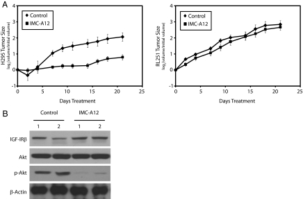
Experimental design: ACC cell lines that display or lack activated IGF signaling are used to assess the effects of two IGF-1R antagonists in cultured cells and ACC xenograft tumors.

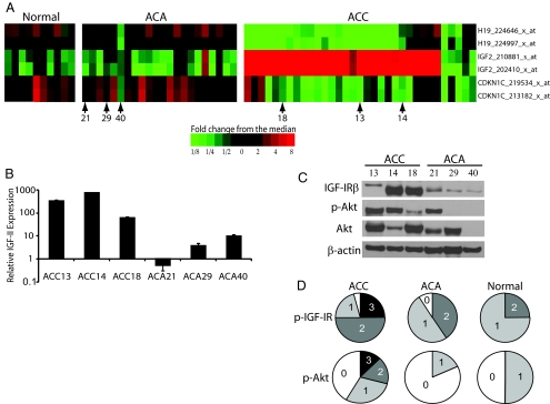
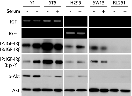
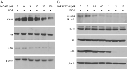
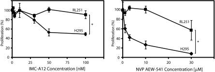
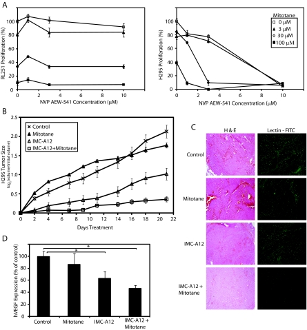
Results: Transcriptional profiling data derived from DNA microarray analysis of human adrenal tumors implicate IGF2 as the single highest up-regulated transcript in the vast majority of carcinomas. We show that the majority of ACC cell lines tested display constitutive IGF ligand production and activation of downstream effector pathways. Both IGF-1R antagonists cause significant dose-dependent growth inhibition in ACC cell lines. Furthermore, we observe that mitotane, the first-line adrenolytic drug used in patients with ACC, results in enhanced growth inhibition when used in combination with the IGF-1R antagonists. We next examined the activity of IGF-1R antagonists against ACC xenografts in athymic nude mice. IGF inhibition markedly reduced tumor growth greater than that observed with mitotane treatment, and combination therapy with mitotane significantly enhanced tumor growth suppression.

Conclusion: These findings establish a critical role of IGF signaling in ACC pathophysiology and provide rationale for use of targeted IGF-1R antagonists to treat adrenocortical carcinoma in future clinical trials.


Figures

Figure 1
Figure 1
Up-regulation of IGF2, overexpression of IGF-1R, and active IGF signaling in human ACCs in comparison with normal and adenoma tissues. A, Snapshot heat map of the 11p15.5 chromosomal region generated from Affymetrix U133A 2.0 Plus oligonucleotide array. Shown are the imprinted genes IGF2, H19, and CDKN1C of 65 patient samples consisting of 10 normal adrenal tissues, 22 adrenal adenomas, and 33 sporadic ACCs. Shades of red indicate increased expression of gene indicated on the right, whereas shades of green reveal decreased expression levels. Numbers below heat map represent location of tumor samples used for quantitative RT-PCR and immunoblotting. B, Quantitative RT-PCR of three ACCs and three adenomas for human IGF2 transcript. The y-axis represents relative IGF2 message levels normalized to glyceraldehyde 3-phosphate dehydrogenase transcript levels. Data shown represent the mean ± sd of triplicate samples of one representative experiment. C, Immunoblot analysis of tumor samples described above for IGF-1R expression, Akt, and activated phosho-AktSer473 as a readout for active IGF signaling. Immunoblotting of β-actin is shown as a protein loading control. D, Histologically graded human tissue microarray staining for phospho-IGF-1R and phospho-Akt with results depicted as percentage pie charts. Immunoreactivity was scored blindly by a four-tier [negative, low (1+), medium (2+), and high (3+) positive] grading scheme of 24 ACCs, 22 ACAs, and four normal adrenal tissue.
Figure 2
Figure 2
Endogenous IGF signaling in a panel of ACC cell lines. A panel of mouse (Y1 and ST5) and human (H295, SW13, and RL251) ACC cell lines cultured in serum-free (−) or serum-containing (+) media assessed for endogenous transcript expression of IGF1 or IGF2 by gel-based RT-PCR (top two panels). Whole-cell lysates were immunoprecipitated (IP) for the IGF-1Rβ and subsequently immunoblotted (IB) for IGF-1Rβ and phospho-tyrosine (middle two panels). Lower two panels show immunoblots from whole-cell lysates for phospho-AktSer473 and total Akt for protein loading control.
Figure 3
Figure 3
IGF-1R antagonist treatments decrease IGF-mediated signaling. A, H295 cells were pretreated with increasing nanomolar concentrations of IMC-A12 for 1 h before addition of 10 nm IGF-I/II ligand mix for 10 min. Cells were subsequently harvested and immunoblotted (IB) for IGF-1Rβ, Akt, phospho-AktSer473, or β-actin. B, H295 cells were pretreated with increasing micromolar concentrations of NVP-AEW541 for 30 min before addition of 10 nm IGF-I/II ligand mix for 10 min. Cells were then harvested and either immunopreciptated (IP) with an anti-IGF-1Rβ antibody and immunoblotted for phospho-tyrosine residues or directly immunoblotted for IGF-1Rβ, Akt, phospho-AktSer473, or β-actin.
Figure 4
Figure 4
Antiproliferative effects of IGF-1R antagonist treatments in vitro. H295 and RL251 cells were incubated with increasing micromolar concentrations of NVP-AEW541 (right panel) or nanomolar concentrations of IMC-A12 (left panel), and proliferation was assessed with MTS reagent. Squares (▪) represent the mean of quadruplicate wells of RL251 cells, whereas diamonds (♦) represent quadruplicate wells of H295 cells with error bars indicating ± sd. Data are representative of at least four independent experiments. *, P < 0.05.
Figure 5
Figure 5
Targeted inhibition of tumor growth in vivo. A, H295 (left panel) and RL251 (right panel) cells were injected sc into athymic nude mice and mice were randomized into treatment groups (n = 8 for H295 and n = 10 for RL251). Groups were treated with vehicle or IMC-A12 every other day. Tumor dimensions on control (♦) mice or IMC-A12-treated (▪) mice were measured three times a week for the duration of the 21-d study, and data are shown as the log ratios of tumor size to initial tumor size means ± se. B, To confirm in vivo targeting of IGF inhibition, harvested H295 tumors were lysed and two tumors from each treatment arm were subjected to immunoblotting for IGF-1Rβ, Akt, phospho-AktSer473, and β-actin.
Figure 6
Figure 6
IGF-1R antagonists enhance the inhibitory effects of mitotane. A, RL251 (left panel) and H295 (right panel) cells were incubated in triplicate with a combination of mitotane and increasing concentrations of NVP-AEW541. Proliferation was assessed with MTS reagent. Data are representative of three independent experiments and displayed as mean ± sd. B, Mice harboring H295 xenografts were randomized into four groups (n = 20 per treatment arm) and treated with vehicle or IMC-A12 every other day and/or mitotane once daily for the duration of the experiment. Tumor volumes were measured three times a week. Data are presented as log ratios of tumor size over initial tumor size means ± se. C, Hematoxylin and eosin (H & E)-stained section of H295 tumor xenografts (left panels) at ×40 magnification. Lectin-FITC immunohistochemical analysis (right panels) was performed to detect relative levels of endothelial cells. D, Quantitative RT-PCR of three or four tumors from each treatment arm with primers detecting all four isoforms of human VEGF. The y-axis represents relative VEGF levels normalized to glyceraldehyde 3-phosphate dehydrogenase transcript levels followed by normalization to control tissue values. Each value represents the average of triplicates of two independent experiments and data are presented as the mean ± se. *, P < 0.05.

References

    1. Wajchenberg BL, Albergaria Pereira MA, Medonca BB, Latronico AC, Campos Carneiro P, Alves VA, Zerbini MC, Liberman B, Carlos Gomes G, Kirschner MA 2000 Adrenocortical carcinoma: clinical and laboratory observations. Cancer 88:711–736 - PubMed
    1. Allolio B, Fassnacht M 2006 Clinical review: adrenocortical carcinoma: clinical update. J Clin Endocrinol Metab 91:2027–2037 - PubMed
    1. Icard P, Goudet P, Charpenay C, Andreassian B, Carnaille B, Chapuis Y, Cougard P, Henry JF, Proye C 2001 Adrenocortical carcinomas: surgical trends and results of a 253-patient series from the French Association of Endocrine Surgeons Study Group. World J Surg 25:891–897 - PubMed
    1. Giordano TJ, Thomas DG, Kuick R, Lizyness M, Misek DE, Smith AL, Sanders D, Aljundi RT, Gauger PG, Thompson NW, Taylor JM, Hanash SM 2003 Distinct transcriptional profiles of adrenocortical tumors uncovered by DNA microarray analysis. Am J Pathol 162:521–531 - PMC - PubMed
    1. Velazquez-Fernandez D, Laurell C, Geli J, Hoog A, Odeberg J, Kjellman M, Lundeberg J, Hamberger B, Nilsson P, Backdahl M 2005 Expression profiling of adrenocortical neoplasms suggests a molecular signature of malignancy. Surgery 138:1087–1094 - PubMed

Publication types

MeSH terms