. 2009 Feb;23(2):503-12.
doi: 10.1096/fj.08-118422. Epub 2008 Oct 14.

Hsp90 inhibitor partially corrects nephrogenic diabetes insipidus in a conditional knock-in mouse model of aquaporin-2 mutation

Affiliations

Hsp90 inhibitor partially corrects nephrogenic diabetes insipidus in a conditional knock-in mouse model of aquaporin-2 mutation

Baoxue Yang et al. FASEB J. 2009 Feb.

Abstract

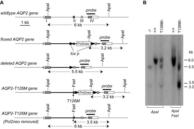
Mutations in aquaporin-2 (AQP2) that interfere with its cellular processing can produce autosomal recessive nephrogenic diabetes insipidus (NDI). Prior gene knock-in of the human NDI-causing AQP2 mutation T126M produced mutant mice that died by age 7 days. Here, we used a novel "conditional gene knock-in" strategy to generate adult, AQP2-T126M mutant mice. Mice separately heterozygous for floxed wild-type AQP2 and AQP2-T126M were bred to produce hemizygous mice, which following excision of the wild-type AQP2 gene by tamoxifen-induced Cre-recombinase gave AQP2(T126M/-) mice. AQP2(T126M/-) mice were polyuric (9-14 ml urine/day) compared to AQP2(+/+) mice (1.6 ml/day) and had reduced urine osmolality (400 vs. 1800 mosmol). Kidneys of AQP2(T126M/-) mice expressed core-glycosylated AQP2-T126M protein in an endoplasmic reticulum pattern. Screening of candidate protein folding "correctors" in AQP2-T126M-transfected kidney cells showed increased AQP2-T126M plasma membrane expression with the Hsp90 inhibitor 17-allylamino-17-demethoxygeldanamycin (17-AAG). 17-AAG increased urine osmolality in AQP2(T126M/-) mice by >300 mosmol but had no effect in AQP2(-/-) mice. Kidneys of 17-AAG-treated AQP2(T126M/-) mice showed partial rescue of defective AQP2-T126M cellular processing. Our results establish an adult mouse model of NDI and demonstrate partial restoration of urinary concentration function by a compound currently in clinical trials for other indications.


Figures

Figure 1.
Figure 1.
Strategy for generation of conditional AQP2-T126M mutant mice. See text for further explanation.
Figure 2.
Figure 2.
Mouse AQP2 gene structure and Southern blot analysis of mutant mice. A) Organization and restriction map of wild-type AQP2 gene (top) and of the various targeted mutant AQP2 genes. Rectangles indicate exon segments, with coding regions shaded. The probe used for Southern blot analysis is indicated (labeled “probe”), along with expected sizes of hybridized fragments after ApaI and/or FseI digestions. B) Southern blot of genomic DNA digested with ApaI and ApaI/FseI with probe as indicated in A.
Figure 3.
Figure 3.
Efficiency of Cre-induced deletion of the floxed wild-type AQP2 gene in AQP2T126M/− mice. A) Left panel: relative copy number of wild-type AQP2 gene in mice of indicated genotypes determined by real-time genomic PCR (mean±se, n=3). Right panel: schematic showing genomic PCR strategy. B) Left panel: relative copy number of mRNA encoding wild-type AQP2 determined by real-time RT-PCR (mean±se, n=3). Right panel: schematic showing RT-PCR strategy. C) Northern blot of kidney mRNA from wild-type and AQP2T126M/− mice probed with the mouse AQP2 cDNA coding sequence. Where indicated, mice were deprived of water for 18 h prior to kidney removal. D) Immunoblot analysis of total mouse kidney protein. Where indicated, protein was digested by endoglycosidase H. Blot probed by polyclonal antibody against the AQP2 C terminus. E) AQP2 immunofluorescence of kidneys of tamoxifen-treated wild-type (left panel) and AQP2T126M/− mice (right panel). White arrowhead indicates a cell expressing wild-type AQP2 protein at the apical plasma membrane.
Figure 4.
Figure 4.
Urinary concentrating function in AQP2T126M/− mice. A) Urine osmolality in AQP2T126M/− mice given free access to food and water (mean±se, 3 mice/group). Arrows indicate tamoxifen (4OH-TM) injections. B) Twenty-four-hour urine output in wild-type mice, (tamoxifen-treated) AQP2T126M/− mice, and AQP2−/− mice. C) Urine osmolality before (H) and after (D) 18-h water deprivation. D) Body weight loss for the same mice in C after water deprivation. E) Left: relative expression of mRNAs encoding wild-type and T126M-AQP2 measured by real-time RT-PCR. Right: upstream PCR primers indicated (underlined) in partial sequence of wild-type and T126M mutant AQP2 cDNA.
Figure 5.
Figure 5.
Partial correction of defective urinary concentrating function in AQP2T126M/− by 17-AAG. A) AQP2 immunoblot of cell surface protein (following biotinylation and avidin column purification) and total protein from MDCK cells expressing wild-type AQP2 or AQP2-T126M. Where indicated, cells were treated for 20 h with 2 μM of 17-AAG, 17-DMAG, geldanamycin, or Congo red. Arrow indicates increased cell surface AQP2-T126M in 17-AAG-treated cells. B) Urine osmolality in AQP2T126M/− mice receiving intraperitoneal injections of 17-AAG (50 mg/kg) or DMSO vehicle (mean±se, 6 mice/group). C) Urine osmolality in wild-type, AQP2T126M/−, and AQP2−/− mice before and after 17-AAG treatment (mean±se, 4 mice/group).
Figure 6.
Figure 6.
Partial rescue of defective AQP2-T126M cellular processing by 17-AAG in kidneys of AQP2T126M/− mice. A) Top: AQP2 immunoblot of kidney homogenates from wild-type and AQP2T126M/− mice. Mice were fully treated (as in Fig. 5A) with 17-AAG were indicated. Arrow indicates the corrected AQP2-T126M protein. Bottom: averaged total AQP2 protein (se, open bars) and core glycosylation protein (solid bars). *P < 0.05 vs. total AQP2 protein from untreated AQP2T126M/− mice. **P < 0.01 vs. core-glycosylated AQP2 protein from untreated AQP2T126M/− mice. B) AQP2 immunofluorescence of kidneys from wild-type (left) and AQP2T126M/− mice, without (middle) or following (right) 17-AAG treatment. Confocal imaging done at two magnifications.

References

    1. Deen P M, Knoers N V. Physiology and pathophysiology of the aquaporin-2 water channel. Curr Opin Nephrol Hypertens. 1998;7:37–42. - PubMed
    1. Nielsen S, Frøkiaer J, Marples D, Kwon T H, Agre P, Knepper M A. Aquaporins in the kidney: from molecules to medicine. Physiol Rev. 2002;82:205–244. - PubMed
    1. Brown D. The ins and outs of aquaporin-2 trafficking. Am J Physiol Renal Physiol. 2003;284:F893–F901. - PubMed
    1. Noda Y, Sasaki S. Trafficking mechanism of water channel aquaporin-2. Biol Cell. 2005;97:885–892. - PubMed
    1. Verkman A S. Dissecting the roles of aquaporins in renal pathophysiology using transgenic mice. Semin Nephrol. 2008;28:217–226. - PMC - PubMed

Publication types

LinkOut - more resources