Review
. 2008 Oct;21(4):639-65.
doi: 10.1128/CMR.00022-08.

New insights on classification, identification, and clinical relevance of Blastocystis spp

Affiliations
Review

New insights on classification, identification, and clinical relevance of Blastocystis spp

Kevin S W Tan. Clin Microbiol Rev. 2008 Oct.

Abstract

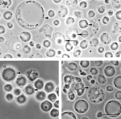
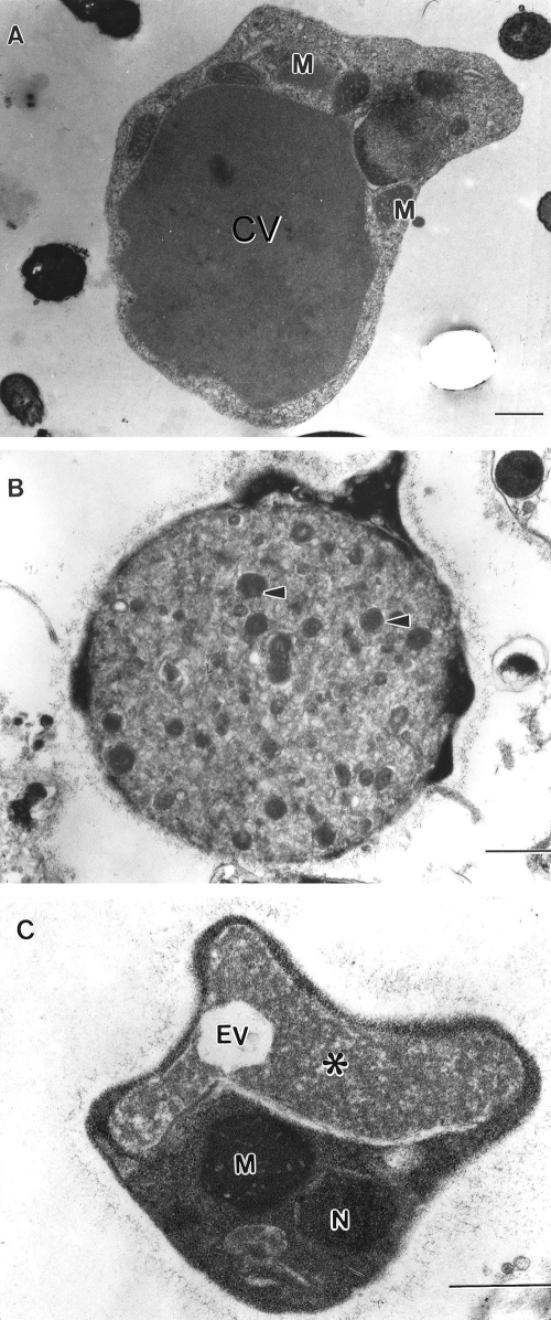
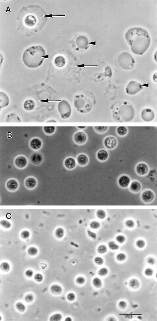
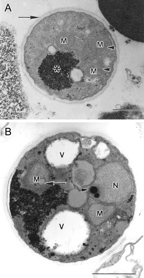
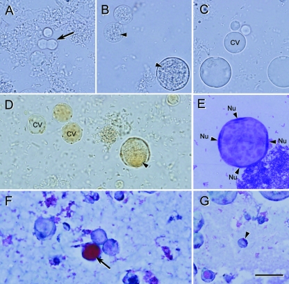
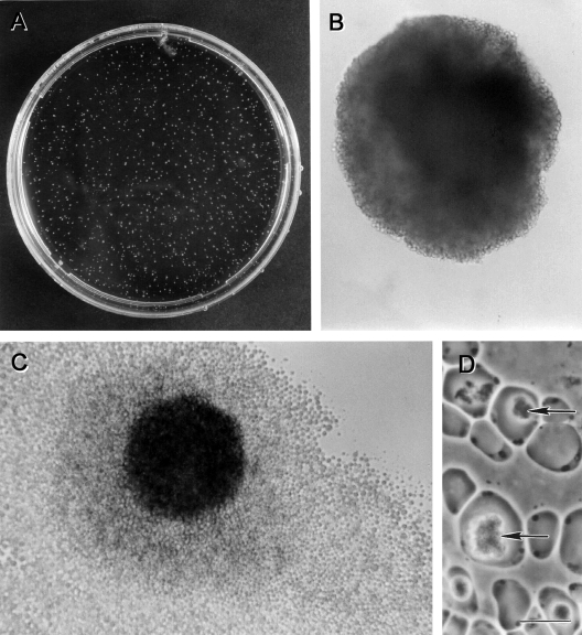
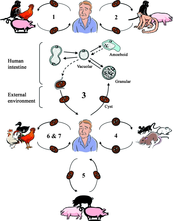
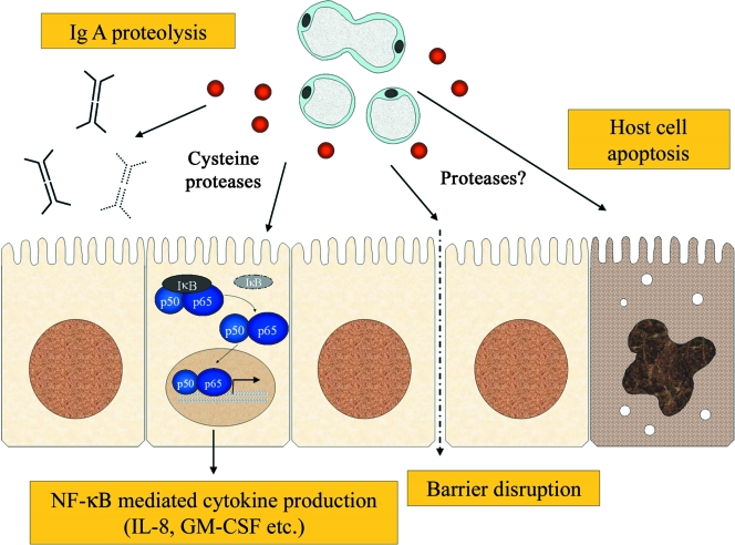
Blastocystis is an unusual enteric protozoan parasite of humans and many animals. It has a worldwide distribution and is often the most commonly isolated organism in parasitological surveys. The parasite has been described since the early 1900s, but only in the last decade or so have there been significant advances in our understanding of Blastocystis biology. However, the pleomorphic nature of the parasite and the lack of standardization in techniques have led to confusion and, in some cases, misinterpretation of data. This has hindered laboratory diagnosis and efforts to understand its mode of reproduction, life cycle, prevalence, and pathogenesis. Accumulating epidemiological, in vivo, and in vitro data strongly suggest that Blastocystis is a pathogen. Many genotypes exist in nature, and recent observations indicate that humans are, in reality, hosts to numerous zoonotic genotypes. Such genetic diversity has led to a suggestion that previously conflicting observations on the pathogenesis of Blastocystis are due to pathogenic and nonpathogenic genotypes. Recent epidemiological, animal infection, and in vitro host-Blastocystis interaction studies suggest that this may indeed be the case. This review focuses on such recent advances and also provides updates on laboratory and clinical aspects of Blastocystis spp.


Figures

FIG. 1.
FIG. 1.
Morphological forms of Blastocystis sp. subtype 4 by phase-contrast microscopy. (A) Vacuolar and fecal cyst forms from in vitro axenic culture displaying extensive size variation (arrowheads). Note the refractile appearance and loose outer coat of cysts (arrows). (B) Granular form with distinct granular inclusions within the central vacuole (arrowhead). (C) Amoeboid forms occasionally seen in culture showing pseudopod-like cytoplasmic extensions (arrow). Bar, 10 μm.
FIG. 2.
FIG. 2.
Transmission electron micrographs of Blastocystis sp. subtype 4. (A) Vacuolar form revealing large central vacuole (CV) resulting in a thin band of peripheral cytoplasm. (B) Granular form revealing electron-dense granules (arrowheads) occupying the entire central vacuole. Note the surface coat surrounding the parasite. (C) Irregular-shaped amoeboid form with central vacuole (asterisk) and empty vacuole (EV). M, mitochondrion-like organelle; N, nucleus. Bar, 1 μm.
FIG. 3.
FIG. 3.
Phase-contrast microscopy of Blastocystis cysts. (A) Spherical cysts of subtype 4 from an in vitro axenic culture displaying a loose outer coat (arrows) among vacuolar forms (arrowheads). (B) Enrichment of subtype 4 cysts and loss of outer coat are apparent after overnight incubation in distilled water. (C) Cysts from a human isolate revealing ovoid morphology distinct from the spherical cysts of subtype 4. Bar, 10 μm.
FIG. 4.
FIG. 4.
Transmission electron micrographs of Blastocystis sp. subtype 4 cysts. (A) Mature cyst with a distinct double-layered cyst wall (arrow) and reduced glycogen mass (asterisk). Mitochondrion-like organelles (M) contain faint saccate cristae (arrowhead). (B) Mature cyst containing large vacuoles (V), a nucleus (N) with a dense chromatin mass (arrowhead), and mitochondrion-like organelles (M) with saccate and circular (arrow) cristae. Bar, 1 μm.
FIG. 5.
FIG. 5.
Light microscopy of nonaxenic Blastocystis sp. subtype 1 cultured in Jones’ medium. (A to C) Unstained wet mounts of various diagnostic forms of Blastocystis. (A) Cells undergoing binary fission (arrow). (B) Granular forms commonly seen in laboratory culture. (C) Vacuolar forms with virtually indiscernible thin cytoplasmic rims. (D) Iodine-stained wet mount revealing cells with distinct organelles, a cytoplasmic rim, and granular inclusions (arrowhead). (E) Giemsa-stained permanent smear of the large vacuolar form containing four nuclei (Nu) evenly distributed around the cytoplasmic rim. (F and G) Trichrome-stained permanent smear of vacuolar forms. The central vacuole of some cells stain strongly (arrow), while others stain less intensely; this may be due to the biochemical heterogeneity of the vacuolar contents. (G) Small vacuolar form (arrowhead) revealing organelles within the cytoplasmic rim. Bar, 20 μm.
FIG. 6.
FIG. 6.
Colonies of Blastocystis sp. subtype 4 after 12 days of culture on an agar plate containing 0.3% Bacto agar in Iscove's modified Dulbecco's medium containing 10% horse serum. (A) Petri dish with numerous buff-colored colonies. (B and C) Magnified view of single colonies. (C) Occasionally, parasite cells spread out at the interface of the agar and the bottom surface of the plate. (D) Amoeboid cells isolated from a colony with large inclusions (arrows) within the central vacuole. Bar, 10 μm.
FIG. 7.
FIG. 7.
Proposed life cycle for Blastocystis cells taking into account recent studies (163, 169, 201, 295) suggesting the existence of zoonotic genotypes (subtypes 1 to 7) with various host specificities. Humans and animals are infected by fecal cysts, which develop into vacuolar forms in the large intestines. In humans, vacuolar forms divide by binary fission and may develop into amoeboid or granular forms. Vacuolar forms undergo encystation in the host intestines, and intermediate cyst forms may be surrounded by a thick fibrillar layer that is subsequently lost during passage in the external environment. Information on the transition from the amoeboid to the vacuolar form and from the vacuolar to the cyst form is lacking. These hypothetical pathways are represented by dotted lines. Subtype 1 is cross-infective among mammalian and avian isolates; subtypes 2, 3, 4, and 5 comprise primate/pig, human, cattle/pig, and rodent isolates, respectively; and subtypes 6 and 7 include avian isolates. The proposed scheme suggests that humans are potentially infected by seven or more species of Blastocystis and that certain animals represent reservoirs for transmission to humans. (Adapted from reference with permission from Taylor and Francis.)
FIG. 8.
FIG. 8.
Model for pathogenesis of Blastocystis spp. Blastocystis infection may result in a variety of pathological outcomes such as secretory IgA degradation, barrier function compromise, host cell apoptosis, and induction of proinflammatory cytokines. IgA degradation and barrier disruption may promote the growth and invasion of neighboring pathogens. GM-CSF, granulocyte-macrophage colony-stimulating factor. (Adapted from reference with permission from Taylor and Francis.)

Similar articles

Cited by

References

    1. Abe, N. 2004. Molecular and phylogenetic analysis of Blastocystis isolates from various hosts. Vet. Parasitol. 120:235-242. - PubMed
    1. Abe, N., Z. Wu, and H. Yoshikawa. 2003. Molecular characterization of Blastocystis isolates from birds by PCR with diagnostic primers and restriction fragment length polymorphism analysis of the small subunit ribosomal RNA gene. Parasitol. Res. 89:393-396. - PubMed
    1. Abe, N., Z. Wu, and H. Yoshikawa. 2003. Molecular characterization of Blastocystis isolates from primates. Vet. Parasitol. 113:321-325. - PubMed
    1. Abe, N., Z. Wu, and H. Yoshikawa. 2003. Zoonotic genotypes of Blastocystis hominis detected in cattle and pigs by PCR with diagnostic primers and restriction fragment length polymorphism analysis of the small subunit ribosomal RNA gene. Parasitol. Res. 90:124-128. - PubMed
    1. Abou El Naga, I. F., and A. Y. Negm. 2001. Morphology, histochemistry and infectivity of Blastocystis hominis cyst. J. Egypt. Soc. Parasitol. 31:627-635. - PubMed

Publication types

MeSH terms

LinkOut - more resources