. 2008 Dec;180(4):2081-94.
doi: 10.1534/genetics.108.095141. Epub 2008 Oct 14.

Drosophila asterless and vertebrate Cep152 Are orthologs essential for centriole duplication

Affiliations

Drosophila asterless and vertebrate Cep152 Are orthologs essential for centriole duplication

Stephanie Blachon et al. Genetics. 2008 Dec.

Abstract

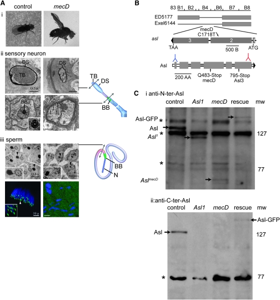
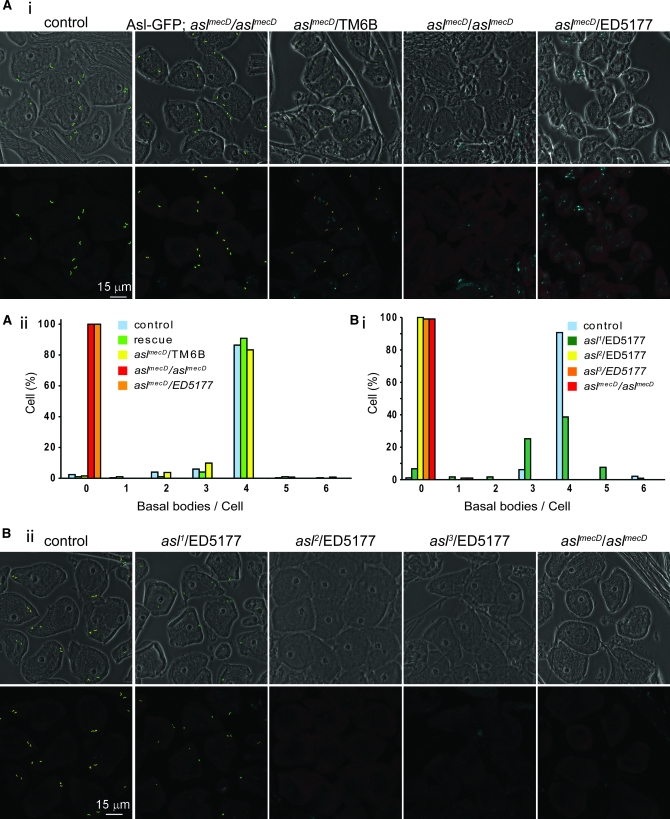
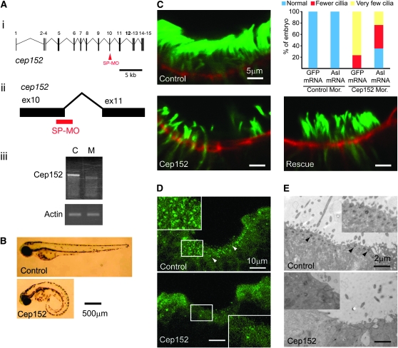
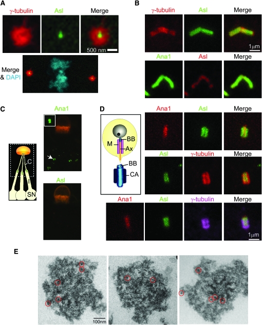
The centriole is the core structure of centrosome and cilium. Failure to restrict centriole duplication to once per cell cycle has serious consequences and is commonly observed in cancer. Despite its medical importance, the mechanism of centriole formation is poorly understood. Asl was previously reported to be a centrosomal protein essential for centrosome function. Here we identify mecD, a severe loss-of-function allele of the asl gene, and demonstrate that it is required for centriole and cilia formation. Similarly, Cep152, the Asl ortholog in vertebrates, is essential for cilia formation and its function can be partially rescued by the Drosophila Asl. The study of Asl localization suggests that it is closely associated with the centriole wall, but is not part of the centriole structure. By analyzing the biogenesis of centrosomes in cells depleted of Asl, we found that, while pericentriolar material (PCM) function is mildly affected, Asl is essential for daughter centriole formation. The clear absence of several centriolar markers in mecD mutants suggests that Asl is critical early in centriole duplication.


Figures

F<sc>igure</sc> 1.—
Figure 1.—
Asl is required for cilia formation. (A) Ciliary defect in mecD mutants. (i) In contrast to control animals, mecD flies cannot stand on their legs, which are crossed, and their wings extend upward. (ii) EM cross sections of the cilia of mechanosensory neurons; in contrast to the control, in the mutants the dendrite tip marked by the dendritic sheath (DS) lacks the tubular body (TB) (top). At the base of sensory cilium (bottom), control but not mecD flies have basal bodies (BB) (inset). In mecD flies, only fragments of the tubular body (arrows) are found. (iii) EM cross sections of spermatid tails; the axoneme (arrows) is missing in the mutant and mitochondrial derivatives (arrowheads) are abnormal (top). Inset shows higher magnification of mitochondrial derivatives and axoneme. A Z-projection of a spermatid cyst where basal bodies labeled by GFP–PACT (arrowhead and inset) are sitting below the nuclei (DAPI, blue) in control and are absent in mutants (bottom). (B) Genetic map illustrating the chromosomal deficiencies (indicated by thin lines) that do not complement aslmecD. The aslmecD mutation introduces a C-to-T transformation near the end of the second exon. aslmecD mutation transforms Q 483 in the fourth coiled-coil domain (shaded squares) to an early stop codon. For comparison, the stop position in asl3 is indicated: in blue, the recognition site of the N-terminal antibody (2891), and in red, the recognition site of the C-terminal antibody (AP1193). (C) Endogenous and GFP fusion proteins are well recognized by both antibodies. The N-terminal antibody (i) detects a faint band of the short Asl protein in aslmecD and a modest level of a larger protein in asl1. Nothing is detected in mutants with the C-terminal antibody (ii). Asterisk is a nonspecific band.
F<sc>igure</sc> 2.—
Figure 2.—
Allelic series analysis of Asl mutations (A) (i) Phase contrast (top) and fluorescent (bottom) photos show that aslmecD homozygous mutants and hemizygotes are devoid of basal bodies whereas heterozygotes, rescue, and control flies have a similar number of centrioles. (ii) Quantification of the number of basal bodies per cell for one representative experiment; for each genotype, ∼100 cells have been counted and two to three flies have been checked. (B) (i) Ana1-GFP labeling shows that basal bodies are present in asl1 and control spermatocytes but are absent in asl2,3 and mecD. Graph shows quantification of basal bodies per cell; for each genotype, ∼100 cells have been counted (ii). Similar results were obtained using Ana1 antibody (data not shown).
F<sc>igure</sc> 3.—
Figure 3.—
Asl function is conserved in zebrafish. (A) Anti-Cep152 morpholino treatment produces a shorter product that lacks 92 bp and contains a frameshift. (i and ii) Location of morpholino target site (red) in the zebrafish Cep152 gene. Gene structure is predicted largely on the basis of publicly available genomic data. (iii) RT–PCR amplification of the Cep152 or Actin transcript at 24 hr post-fertilization in embryos treated with a control (C) or anti-Cep152 morpholino (M). (B) Curly tail, a phenotype characteristic of ciliary defects, is observed in Cep152 morphant larvae. (C) Quantification of cilia number in olfactory pits of morphant embryos. GFP or Asl mRNA are injected along with a control (control mor.) or anti-Cep152 splice site morpholino (Cep152 mor.). Anti-acetylated tubulin antibody (green) was used to stain cilia, and phaloidin (red) marks the apical surface of the tissue. Colored bars indicate the relative quantities of embryos in three phenotypic categories: blue, 50–100% of the wild-type cilia number; red, 25–50%; and yellow, <25%. For each group, n ≥ 10. (D) Staining of basal bodies (arrowheads) in the nasal pits of control and Cep152 morphant embryo with antibodies to γ-tubulin. Insets show higher magnification of areas in white boxes. (E) The apical surface of nasal pits in control and Cep152 morphant embryos visualized by electron microscopy. Basal bodies are indicated with arrowheads. Insets show higher magnification of this tissue.
F<sc>igure</sc> 4.—
Figure 4.—
Asl is a pericentriolar protein closely associated with the centriole wall. (A) In the early embryo, Asl-GFP localizes to the center of the centrosome. The mitotic PCM is marked by γ-tubulin. DAPI stains chromosomes. (B) In spermatocytes, Asl-GFP (top) and endogenous Asl labeled with antibody (bottom) mark the basal body along its length. (C) Unlike the centriolar protein Ana1, Asl is not a component of the basal body of sensory neurons (arrow). The cuticular structure is highlighted by auto-fluorescence (orange). Inset: zoom on basal body. Diagram depicting the location of the basal body (green) and cilia (C) in the sensory organ (SN). (D) Diagram depicting in early spermatids the PCM/centriolar adjunct (CA) surrounding the basal body (BB). M, mitochondria, Ax, axoneme. Asl-GFP (green) is localized to the centriolar adjunct surrounding the basal body marked by Ana1-tdTomato (red) (top). γ-Tubulin staining labels the centriolar adjunct but is even more peripheral than Asl (middle and bottom). (E) Immuno-EM of centrosomes purified from fly embryos expressing Asl-GFP; the 5-nm gold particles are circled in red. Dashed lines indicate the border of the centriole.
F<sc>igure</sc> 5.—
Figure 5.—
Asl is essential for daughter centriole formation. (A) In control metaphase cells, the centriolar marker GFP–PACT colocalizes with γ-tubulin and Cnn at the spindle pole (12 of the 12 cells analyzed). In aslmecD, chromosomes (DAPI) are normal but centrosomes are absent (8 of the 8 cells analyzed). (B) Asl is not essential for centriole maintenance. In wild-type testes, cells are filled with centrioles (Ana1-GFP, green) while in aslmecD a population of MC centrioles (green) is found at the tip of the testes (white circle) marked with anti-FasIII (red). DAPI-stained DNA is blue; insets provide magnification of centrioles. Graph below shows quantification of the number of centrioles in the tip of the testes at different stages of development. (C) EM sections of the tip of the testis show a pair of mother (M) and daughter (D) centrioles as control and a single centriole (M) in aslmecD testis. (D) Spermatocytes expressing Ana1-GFP (green) and stained for γ-tubulin (red) show mother and daughter centrioles in control as a 𝒱-shape. In aslmecD, γ-tubulin staining is normal but only one centriole can be seen. The basal body length is similar in control and aslmecD (graph below).
F<sc>igure</sc> 6.—
Figure 6.—
Maternally contributed centrioles: a method for studying centriolar protein function. (A) In the embryos of flies, a stock of wild-type proteins is provided by the heterozygous mother until transcription starts. It allows for the formation of a certain number of wild-type centrioles: MC centrioles (in green). Polar cells (red) localized in the posterior part of the embryos contain MC centrioles and give rise to germ stem cells. (B and C) Polar cells become the germline stem cells of the larva and the pupa. In pupae, they are localized at the tip of the testis and can be stained with anti-fasIII antibody (E and Figure 5B). (D) In stem cells, the mother centriole (green) stays attached to the cell while the daughter centriole (blue) is inherited by differentiating cells. In mutants, we can ask if MC centrioles are maintained in the stem cells. We can study the fate of the daughter centrioles (blue) during differentiation and ask if they are capable of duplicating or elongating. Are they stable and conserve the same size during time? Are they able to recruit PCM? While new centrioles are not formed after Asl depletion, the fate of the few daughter centrioles formed during the time of maternal contribution can be analyzed by dissecting animals at larval, early, and late pupal stages. (E) In aslmecD, MC centrioles (green) stay in the tip of the testis marked by fasIII antibody (red) from larval to dark pupal stages. In contrast, in spermatocytes some MC centrioles are present in larvae, but at the pupal stage they have disappeared as the spermatocytes that had a centriole in larvae have differentiated into spermatids. (F) During spermatocyte growth, centrioles (C) duplicate and elongate (diagram). Spermatocytes at different stages stained by anti-Cter-Asl (AP1193) show absence of Asl on MC centrioles in aslmecD.
F<sc>igure</sc> 7.—
Figure 7.—
PCM recruitment is not impaired in aslmecD. (A) In aslmecD, quantification of the total intensity of the γ-tubulin (red) signal per centriole shows a significant reduction in mature spermatocytes but normal amounts in meiosis. In the graphs (right), green indicates the intensity of Ana1-GFP used as a control and gray the intensity of the background. (B) In contrast and as previously reported, asl1 shows a dramatic reduction of the total intensity of γ-tubulin signal per centriole in meiosis.
F<sc>igure</sc> 8.—
Figure 8.—
asl1 accumulates a normal amount of PCM in mitosis. (A) Spermatogonia in mitosis of asl1 hemizygotes are able to assemble PCM. Quantification of the total intensity of γ-tubulin signal shows a slight reduction in asl1compared with control. (B) Mitotic cells in larval brains of asl1 homozygotes accumulate normal amounts of PCM. However, half of the cells contain fewer than two centrosomes. Like aslmecD mutants (Figure 5A), asl2 and asl3 are devoid of centrosomes. (C) In asl1 mutant testis expressing Ana1-GFP, the anti-N-terminal Asl antibody detects the Asl1 protein on centrioles in young spermatocytes, but shows abnormal localization of Asl1 to the distal part of the basal body in mature spermatocytes. MC centrioles present in asl3 larval testis are not stained by anti-Asl.

Similar articles

Cited by

References

    1. Andersen, J. S., C. J. Wilkinson, T. Mayor, P. Mortensen, E. A. Nigg et al., 2003. Proteomic characterization of the human centrosome by protein correlation profiling. Nature 426 570–574. - PubMed
    1. Anderson, R. G., and R. M. Brenner, 1971. The formation of basal bodies (centrioles) in the Rhesus monkey oviduct. J. Cell Biol. 50 10–34. - PMC - PubMed
    1. Avidor-Reiss, T., A. M. Maer, E. Koundakjian, A. Polyanovsky, T. Keil et al., 2004. Decoding cilia function: defining specialized genes required for compartmentalized cilia biogenesis. Cell 117 527–539. - PubMed
    1. Basto, R., J. Lau, T. Vinogradova, A. Gardiol, C. G. Woods et al., 2006. Flies without centrioles. Cell 125 1375–1386. - PubMed
    1. Bonaccorsi, S., M. G. Giansanti and M. Gatti, 1998. Spindle self-organization and cytokinesis during male meiosis in asterless mutants of Drosophila melanogaster. J. Cell Biol. 142 751–761. - PMC - PubMed

Publication types

MeSH terms