

Co-expression of KLK6 and KLK10 as prognostic factors for survival in pancreatic ductal adenocarcinoma
- PMID: 18854834
- PMCID: PMC2579692
- DOI: 10.1038/sj.bjc.6604717
Co-expression of KLK6 and KLK10 as prognostic factors for survival in pancreatic ductal adenocarcinoma
Abstract
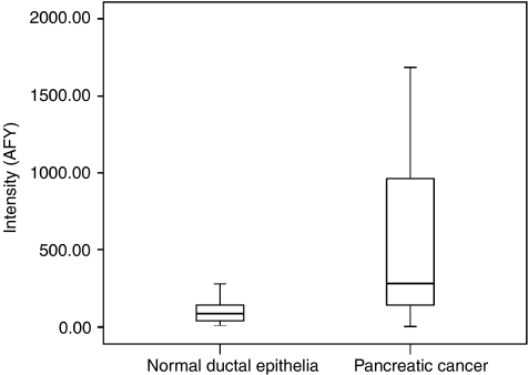
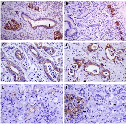
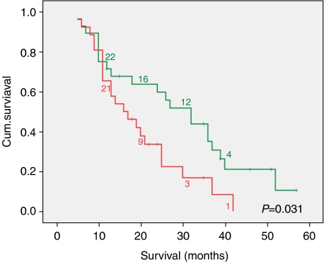
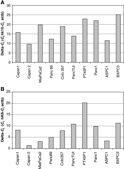
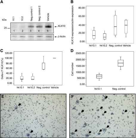
Kallikreins play an important role in tumour microenvironment and as cancer biomarkers in different cancer entities. Previous studies suggested an upregulation of KLK10 and KLK6 in pancreatic ductal adenocarcinoma (PDAC). Therefore, we evaluated the clinicopathological role of these kallikreins and their value as biomarkers in PDAC.Differential expression was validated by DNA-microarrays and immunohistochemistry in normal and malignant pancreatic tissues. Sera concentrations of both kallikreins were evaluated using ELISA. In silico analysis of possible protein interactions and gene silencing of KLK10 in vitro using siRNAs gave further insights in the pathomechanisms.Gene expression analysis and immunohistochemistry demonstrated a strong expression for KLK10 and KLK6 in PDAC. Statistical analysis showed that co-expression of these kallikreins correlated with an R1-resection status (P=0.017) and worse outcome for overall survival (P=0.031). Multivariate analysis proofed that co-expression is an independent prognostic factor for survival (P=0.043). Importantly, KLK10 knockdown in AsPC-1 cells significantly reduced cell migration, whereas computational analysis suggested interaction of KLK6 with angiogenetic factors as an important mechanism.Co-expression of KLK10 and KLK6 plays an unfavourable role in PDAC. Our results suggest that this effect is likely mediated by an interaction with the factors of the extracellular matrix and enhancement of cancer cell motility.
Figures
Similar articles
-
In-silico analysis of kallikrein gene expression in pancreatic and colon cancers.Anticancer Res. 2004 Jan-Feb;24(1):43-51. Anticancer Res. 2004. PMID: 15015574
-
Clinicopathological correlations of nestin expression in surgically resectable pancreatic cancer including an analysis of perineural invasion.J Gastrointestin Liver Dis. 2011 Dec;20(4):389-96. J Gastrointestin Liver Dis. 2011. PMID: 22187705
-
Aberrant upregulation of KLK10 promotes metastasis via enhancement of EMT and FAK/SRC/ERK axis in PDAC.Biochem Biophys Res Commun. 2018 May 15;499(3):584-593. doi: 10.1016/j.bbrc.2018.03.194. Epub 2018 Apr 4. Biochem Biophys Res Commun. 2018. PMID: 29621546
-
Low RASSF6 expression in pancreatic ductal adenocarcinoma is associated with poor survival.World J Gastroenterol. 2015 Jun 7;21(21):6621-30. doi: 10.3748/wjg.v21.i21.6621. World J Gastroenterol. 2015. PMID: 26074700 Free PMC article.
-
Pancreatic stellate cells and pancreas cancer: current perspectives and future strategies.Eur J Cancer. 2014 Oct;50(15):2570-82. doi: 10.1016/j.ejca.2014.06.021. Epub 2014 Aug 1. Eur J Cancer. 2014. PMID: 25091797 Review.
Cited by
-
Identification of human tissue kallikrein 6 as a potential marker of laryngeal cancer based on the relevant secretory/releasing protein database.Dis Markers. 2014;2014:594093. doi: 10.1155/2014/594093. Epub 2014 Feb 11. Dis Markers. 2014. PMID: 24669031 Free PMC article.
-
Cysteine depletion induces pancreatic tumor ferroptosis in mice.Science. 2020 Apr 3;368(6486):85-89. doi: 10.1126/science.aaw9872. Science. 2020. PMID: 32241947 Free PMC article.
-
KLK10 exon 3 unmethylated PCR product concentration: a new potential early diagnostic marker in ovarian cancer? - A pilot study.J Ovarian Res. 2018 Apr 24;11(1):32. doi: 10.1186/s13048-018-0407-y. J Ovarian Res. 2018. PMID: 29690914 Free PMC article.
-
Identification of Prognostic Biomarkers and Drugs Targeting Them in Colon Adenocarcinoma: A Bioinformatic Analysis.Integr Cancer Ther. 2019 Jan-Dec;18:1534735419864434. doi: 10.1177/1534735419864434. Integr Cancer Ther. 2019. PMID: 31370719 Free PMC article.
-
A 65‑gene signature for prognostic prediction in colon adenocarcinoma.Int J Mol Med. 2018 Apr;41(4):2021-2027. doi: 10.3892/ijmm.2018.3401. Epub 2018 Jan 18. Int J Mol Med. 2018. PMID: 29393333 Free PMC article.
References
-
- Bernett MJ, Blaber SI, Scarisbrick IA, Dhanarajan P, Thompson SM, Blaber M (2002) Crystal structure and biochemical characterization of human kallikrein 6 reveals that a trypsin-like kallikrein is expressed in the central nervous system. J Biol Chem 277: 24562–24570 - PubMed
-
- Borgono CA, Diamandis EP (2004) The emerging roles of human tissue kallikreins in cancer. Nat Rev Cancer 4: 876–890 - PubMed
-
- Borgono CA, Michael IP, Diamandis EP (2004) Human tissue kallikreins: physiologic roles and applications in cancer. Mol Cancer Res 2: 257–280 - PubMed
-
- Dawelbait G, Winter C, Zhang Y, Pilarsky C, Grutzmann R, Heinrich JC, Schroeder M (2007) Structural templates predict novel protein interactions and targets from pancreas tumour gene expression data. Bioinformatics 23: i115–i124 - PubMed
Publication types
MeSH terms
Substances
LinkOut - more resources
Full Text Sources
Other Literature Sources
Medical
