Review
. 2009 Jan;335(1):17-25.
doi: 10.1007/s00441-008-0694-5. Epub 2008 Oct 15.

Organization and signaling of endothelial cell-to-cell junctions in various regions of the blood and lymphatic vascular trees

Affiliations
Review

Organization and signaling of endothelial cell-to-cell junctions in various regions of the blood and lymphatic vascular trees

Elisabetta Dejana et al. Cell Tissue Res. 2009 Jan.

Abstract

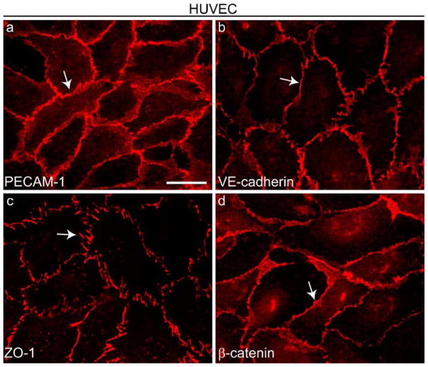
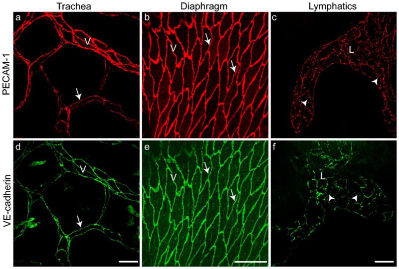
Adhesive intercellular junctions between endothelial cells are formed by tight junctions and adherens junctions. In addition to promoting cell-to-cell adhesion, these structures regulate paracellular permeability, contact inhibition of endothelial cell growth, cell survival, and maintenance of cell polarity. Furthermore, adherens junctions are required for the correct organization of new vessels during embryo development or during tissue proliferation in the adult. Extensive research on cultured epithelial and endothelial cells has resulted in the identification of many molecular components of tight junctions and adherens junctions. Such studies have revealed the complexity of these structures, which are formed by membrane-associated adhesion proteins and a network of several intracellular signaling partners. This review focuses on the structural organization of junctional structures and their functional interactions in the endothelium of blood vessels and lymphatics. We emphasize the way that these structures regulate endothelial cell homeostasis by transferring specific intracellular signals and by modulating activation and signaling of growth factor receptors.


Figures

Fig. 1
Fig. 1
Distribution of cell-to-cell junction proteins in cultured endothelial cells. Cultured human umbilical vein endothelial cells (HUVEC) were grown to confluence and stained by immunofluorescence with antibodies directed against four junctional proteins (arrows): PECAM-1 (a) outside adherens junctions (AJs) and tight junctions (TJs), VE-cadherin (b) at AJs, ZO-1 (c) at TJs and AJs, and β-catenin (d) at AJs. Bar 20 μm
Fig. 2
Fig. 2
Endothelial junction organization in vivo comparable with that observed in cultured endothelial cells (Fig. 1). Staining with antibodies to PECAM-1 (a–c) and VE-cadherin (d–f). The venules (V) of mouse trachea (a, d) and diaphragm (b, e) and the lymphatics (L) of diaphragm (c, f) are immunostained (arrows zipper-like junctions of blood vessels, arrowheads button-like junctions in lymphatics; see also Fig. 3). Bars 50 μm
Fig. 3
Fig. 3
Representation of three types of endothelial junctions (left) and the corresponding electron micrographs (right). In small arterioles (a, b), endothelial cell junctions are tight, probably being formed by TJs intermingled with AJs to limit exchange between blood and tissues. In venules (c, d), junctions are formed by AJs, and small areas of TJs are frequently concentrated at the apical side of the intercellular cleft. In initial lymphatics, (e, f) in which intercellular junctions control entry (intravasation) and drainage of fluid and cells from tissues, junctions are permeable, and endothelial borders have discontinuous button-like junctions with intermingled flaps resembling valve-like structures (the junctional structures are presented en face; from Baluk et al. 2007). AJ and TJ proteins are concentrated at the buttons and allow the flaps to open freely without disrupting overall vascular organization. The more distal collecting lymphatic vessels have continuous zipper-like junctions similar to those of blood vessels (not shown). In addition to AJ and TJ proteins, other junctional adhesive proteins and intracellular partners are present at endothelial junctions but, for simplicity, are not reported here. Note the different scales in the electron micrographs as well as the corresponding drawings. Bars 1 μm

References

    1. Anfosso F, Bardin N, Vivier E, Sabatier F, Sampol J, Dignat-George F. Outside-in signaling pathway linked to CD146 engagement in human endothelial cells. J Biol Chem. 2001;276:1564–1569. - PubMed
    1. Ayalon O, Sabanai H, Lampugnani MG, Dejana E, Geiger B. Spatial and temporal relationships between cadherins and PECAM-1 in cell-cell junctions of human endothelial cells. J Cell Biol. 1994;126:247–258. - PMC - PubMed
    1. Balda MS, Anderson JM. Two classes of tight junctions are revealed by ZO-1 isoforms. Am J Physiol. 1993;264:C918–C924. - PubMed
    1. Baluk P, Fuxe J, Hashizume H, Romano T, Lashnits E, Butz S, Vestweber D, Corada M, Molendini C, Dejana E, McDonald DM. Functionally specialized junctions between endothelial cells of lymphatic vessels. J Exp Med. 2007;204:2349–2362. - PMC - PubMed
    1. Baumeister U, Funke R, Ebnet K, Vorschmitt H, Koch S, Vestweber D. Association of Csk to VE-cadherin and inhibition of cell proliferation. EMBO J. 2005;24:1686–1695. - PMC - PubMed

Publication types

Substances

LinkOut - more resources