. 2009 Jan;217(1):83-93.
doi: 10.1002/path.2445.

The EWSR1/NR4A3 fusion protein of extraskeletal myxoid chondrosarcoma activates the PPARG nuclear receptor gene

Affiliations

The EWSR1/NR4A3 fusion protein of extraskeletal myxoid chondrosarcoma activates the PPARG nuclear receptor gene

C Filion et al. J Pathol. 2009 Jan.

Abstract

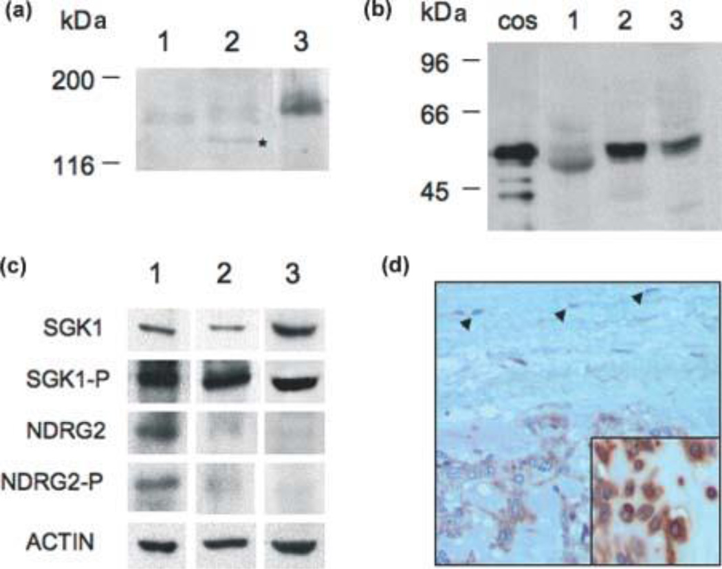
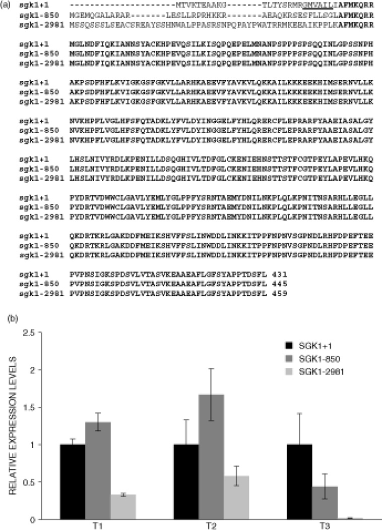
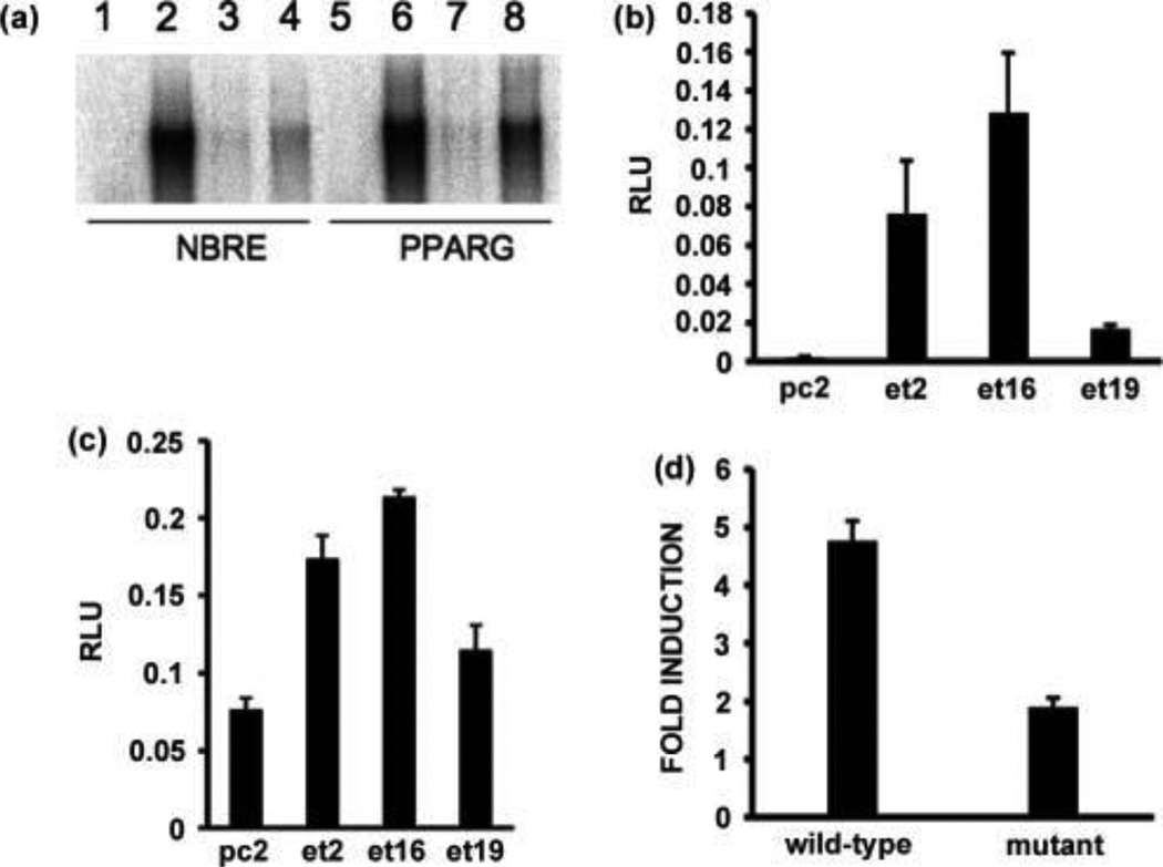
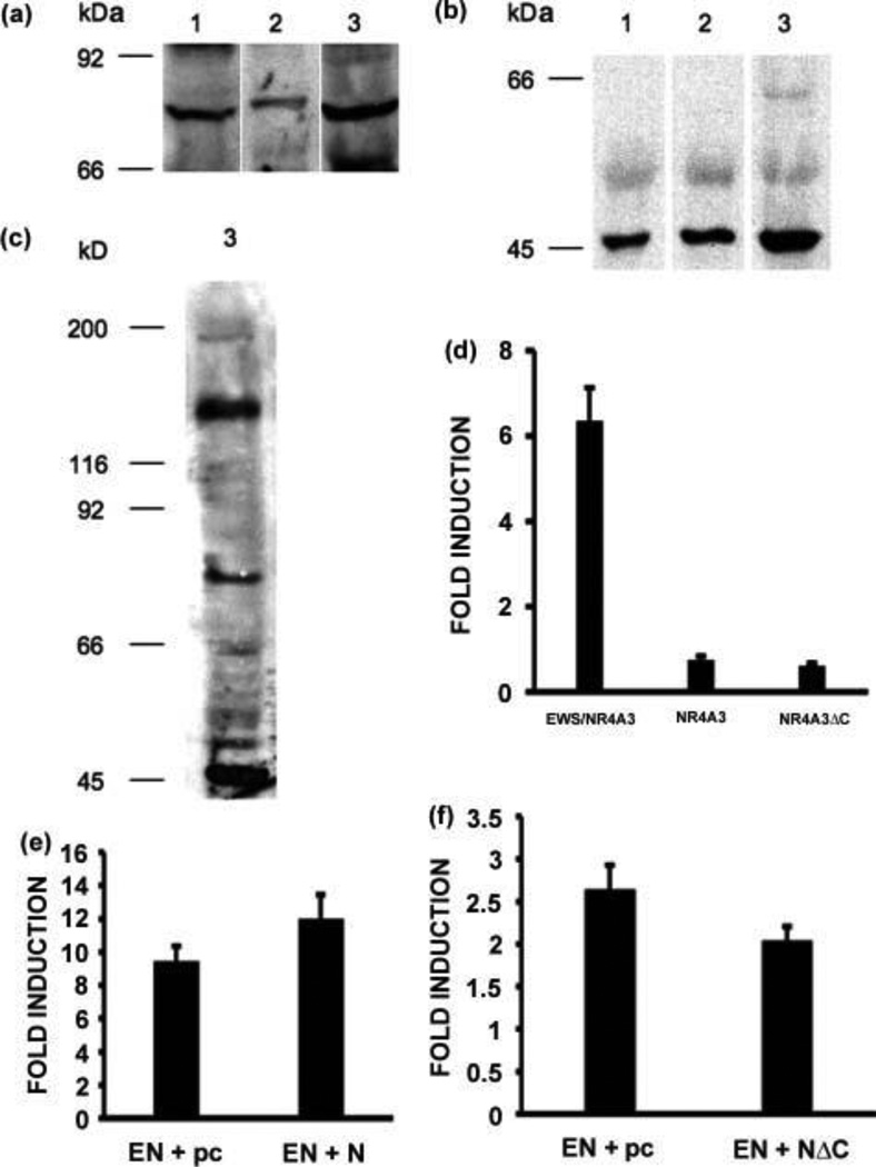
The NR4A3 nuclear receptor is implicated in the development of extraskeletal myxoid chondrosarcoma (EMC), primitive sarcoma unrelated to conventional chondrosarcomas, through a specific fusion with EWSR1 resulting in an aberrant fusion protein that is thought to disrupt the transcriptional regulation of specific target genes. We performed an expression microarray analysis of EMC tumours expressing the EWSR1/NR4A3 fusion protein, comparing their expression profiles to those of other sarcoma types. We thereby identified a set of genes significantly overexpressed in EMC relative to other sarcomas, including PPARG and NDRG2. Western blot or immunohistochemical analyses confirm that PPARG and NDRG2 are expressed in tumours positive for EWSR1/NR4A3. Bioinformatic analysis identified a DNA response element for EWSR1/NR4A3 in the PPARG promoter, and band-shift experiments and transient transfections indicate that EWSR1/NR4A3 can activate transcription through this element. Western blots further show that an isoform of the native NR4A3 receptor lacking the C-terminal domain is very highly expressed in tumours positive for EWSR1/NR4A3, and co-transfections of this isoform along with EWSR1/NR4A3 indicate that it may negatively regulate the activity of the fusion protein on the PPARG promoter. These results suggest that the overall expression of PPARG in EMC may be regulated in part by the balance between EWSR1/NR4A3 and NR4A3, and that PPARG may play a crucial role in the development of these tumours. The specific up-regulation of PPARG by EWSR1/NR4A3 may also have potential therapeutic implications.


Conflict of interest statement

There are no conflicts of interest for any of the authors

Figures

Figure 1
Figure 1
Expression of EWSR1/NR4A3, PPARG, NDRG2 and SGK1 proteins in EMC tumors. (A) Western blot analyses of three EMC tumors demonstrating the expression of the EWSR1/NR4A3 fusion protein using a polyclonal antibody directed against NR4A3 [34]. The lower band detected in tumor sample 2 and marked by a star may correspond to either a degradation product of the full-length fusion protein or a truncated isoform generated by alternative splicing of the fusion mRNA [23]. (B) Western blot analysis of PPARG expression in the same three tumor samples. The lane labeled COS contains a sample of COS cells transfected with a PPARG isoform 1 expression vector. (C) Western blot analysis of NDRG2 and SGK1 proteins in the same three tumor samples. (D) Immunohistochemistry staining for NDRG2 showing diffuse cytoplasmic staining of EMC tumor cells and lack of staining in fibroblasts in fibrotic capsule (arrows) (200×). Inset shows higher magnification (400×) of cytoplasmic staining in another EMC case.
Figure 2
Figure 2
Real-time PCR analyses of SGK1 mRNAs in EMC tumors. (A) Schematic diagram of the three SGK1 protein isoforms encoded by the SGK1+1, SGK1-850 and SGK1-2981 mRNAs. Common amino acids are in bold characters and the proteasomal degradation signal present in the SGK1+1 isoform is underlined. (B) Real-time PCR of the three SGK1 mRNAs in three EMC tumors. For each tumor, a total RNA extract was prepared from which three different reverse-transcription reactions were performed followed by a real-time PCR for each SGK1 mRNA and the 18S ribosomal RNA. The data were normalized with respect to the 18S RNA and the results are expressed as relative expression levels with the SGK1+1 mRNA set at one. The standard deviations are shown as vertical bars.
Figure 3
Figure 3
Activation of the PPARG promoter by EWSR1/NR4A3 through the NBRE site. (A) Band-shift analyses of in vitro translated EWSR1/NR4A3 (lanes 2–4 and 6–8) with NBRE (lanes 1–4) and PPARG (lanes 5–8) oligonucleotides. Lanes 3 and 7 reactions contained a 25-fold excess of cold wild-type oligonucleotides, and lanes 4 and 8 reactions contained a 25 fold excess of mutant oligonucleotides. The reactions in lanes 1 and 5 used a control rabbit reticulocyte lysate sample. (B) and (C) Transient transfections of CFK2 cell lines stably expressing EWSR1/NR4A3 (et2, et16, et19) with a NBRE luciferase reporter vector (B) or a PPARG promoter luciferase reporter vector (C). The pc2 cell line is a negative control cell line stably transfected with an empty expression vector. The results are expressed as relative light units (RLU) and the standard deviation is shown (vertical bars). (D) Transient transfections of wild-type CFK2 cells with an EWSR1/NR4A3 expression vector, a wild-type or mutated PPARG promoter luciferase reporter vector, and a pBIND (Promega) normalization vector. Transfections were carried out in triplicate, the results are expressed as fold induction relative to the empty expression vector, and the standard deviation is shown (vertical bars).
Figure 4
Figure 4
Expression of NR4A3 and NR4A3∆C in EMC tumors. (A), (B) and (C) Western blot analyses of the same three EMC tumors presented in Fig. 1 showing expression of the full-length NR4A3 nuclear receptor (A) and of the truncated NR4A3ΔC isoform (B), and co-expression of EWSR1/NR4A3, NR4A3, and NR4A3ΔC in tumor sample 3 (C). (D), (E) and (F) Transient transfections of CFK2 cells with an EWSR1/NR4A3, NR4A3, or NR4A3∆C expression vector and the PPARG promoter luciferase reporter vector (D), with a five-fold excess of the EWSR1/NR4A3 expression vector compared to the NR4A3 expression vector (E), or with a five-fold excess of the NR4A3∆C expression vector compared to the EWSR1/NR4A3 expression vector (F). Transfections were carried out in triplicate, pBIND (Promega) was used for normalization, results are expressed as fold induction with respect to the empty expression vector, and the standard deviation is shown (vertical bars).

References

    1. Ohkura N, Hijikuro M, Yamamoto A, Miki K. Molecular cloning of a novel thyroid/steroid receptor superfamily gene from cultured rat neuronal cells. Biochemical and biophysical research communications. 1994;205:1959–1965. - PubMed
    1. Milbrandt J. Nerve growth factor induces a gene homologous to the glucocorticoid receptor gene. Neuron. 1988;1:183–188. - PubMed
    1. Hazel TG, Misra R, Davis IJ, Greenberg ME, Lau LF. Nur77 is differentially modified in PC12 cells upon membrane depolarization and growth factor treatment. Molecular and cellular biology. 1991;11:3239–3246. - PMC - PubMed
    1. Scearce LM, Laz TM, Hazel TG, Lau LF, Taub R. RNR-1, a nuclear receptor in the NGFI-B/Nur77 family that is rapidly induced in regenerating liver. The Journal of biological chemistry. 1993;268:8855–8861. - PubMed
    1. Hedvat CV, Irving SG. The isolation and characterization of MINOR, a novel mitogen-inducible nuclear orphan receptor. Molecular endocrinology (Baltimore, Md. 1995;9:1692–1700. - PubMed

Publication types

MeSH terms