

Interstitial adenosine concentration during norepinephrine infusion in isolated guinea pig hearts
- PMID: 1887934
- PMCID: PMC4125619
- DOI: 10.1152/ajpheart.1991.261.3.H901
Interstitial adenosine concentration during norepinephrine infusion in isolated guinea pig hearts
Abstract
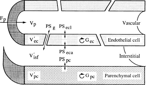
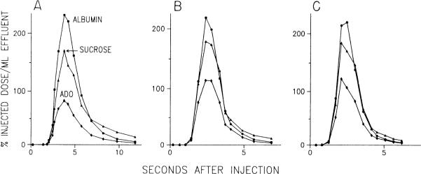
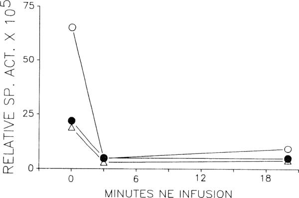
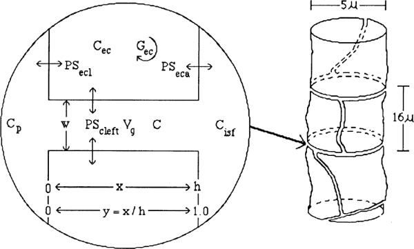
This study determined the effect of norepinephrine (NE) on cardiac interstitial fluid adenosine concentration [( ADO]isf). Isolated guinea pig hearts were perfused with a Krebs-Henseleit buffer solution. Radiolabeled albumin, sucrose, and adenosine were injected under control conditions and after 3 and 20 min of NE infusion to obtain multiple indicator dilution curves that were used to determine capillary transport parameters for adenosine. These parameters together with venous adenosine concentrations were used in a mathematical model to a calculate [ADO]isf. Capillary transport parameters were not changed significantly by NE infusion. Because of uncertainty regarding two model parameters, two sets of [ADO]isf values were calculated. One set used best-fit values obtained from indicator dilution curves, and a second set used parameters chosen to provide the highest [ADO]isf values consistent with indicator dilution curves. Venous adenosine concentrations were 1.9 +/- 0.4 nM under control conditions and 243 +/- 110 and 45 +/- 25 nM after 3 and 20 min of NE infusion, respectively. Calculated [ADO]isf was 2.6-9.4, 591-1,288, and 166-324 nM, respectively, under these same conditions. We conclude that NE infusion greatly increases [ADO]isf, and adenosine is responsible for most of the vasodilation at 3 min. The subsequent fall in venous concentration is due to a fall in [ADO]isf rather than to decreased capillary permeability. Vascular resistance remained low while [ADO]isf fell, which suggests that additional vasodilators are important during maintained NE infusion.
Figures
Similar articles
-
Transcapillary adenosine transport and interstitial adenosine concentration in guinea pig hearts.Am J Physiol. 1989 Jul;257(1 Pt 2):H89-106. doi: 10.1152/ajpheart.1989.257.1.H89. Am J Physiol. 1989. PMID: 2750952 Free PMC article.
-
Cardiac endothelial transport and metabolism of adenosine and inosine.Am J Physiol. 1999 Sep;277(3):H1241-51. doi: 10.1152/ajpheart.1999.277.3.H1241. Am J Physiol. 1999. PMID: 10484446 Free PMC article.
-
Cardiac microdialysis to estimate interstitial adenosine and coronary blood flow.Am J Physiol. 1990 Jun;258(6 Pt 2):H1642-9. doi: 10.1152/ajpheart.1990.258.6.H1642. Am J Physiol. 1990. PMID: 2360661
-
[The receptorial responsiveness method (RRM): a new possibility to estimate the concentration of pharmacologic agonists at their receptors].Acta Pharm Hung. 2014;84(1):38-52. Acta Pharm Hung. 2014. PMID: 24809165 Review. Hungarian.
-
Indicator dilution estimation of capillary endothelial transport.Annu Rev Physiol. 1986;48:321-34. doi: 10.1146/annurev.ph.48.030186.001541. Annu Rev Physiol. 1986. PMID: 3518617 Free PMC article. Review.
Cited by
-
Transient transcapillary exchange of water driven by osmotic forces in the heart.Am J Physiol Heart Circ Physiol. 2003 Sep;285(3):H1317-31. doi: 10.1152/ajpheart.00587.2002. Epub 2003 May 8. Am J Physiol Heart Circ Physiol. 2003. PMID: 12738617 Free PMC article.
-
Multiscale modeling of cardiac cellular energetics.Ann N Y Acad Sci. 2005 Jun;1047:395-424. doi: 10.1196/annals.1341.035. Ann N Y Acad Sci. 2005. PMID: 16093514 Free PMC article. Review.
-
Myocardial adenosine stimulates release of cyclic adenosine monophosphate from capillary endothelial cells in guinea pig heart.Pflugers Arch. 1993 May;423(3-4):330-7. doi: 10.1007/BF00374413. Pflugers Arch. 1993. PMID: 8391684
-
Strategies and Tactics in Multiscale Modeling of Cell-to-Organ Systems.Proc IEEE Inst Electr Electron Eng. 2006 Apr;94(4):819-830. doi: 10.1109/JPROC.2006.871775. Proc IEEE Inst Electr Electron Eng. 2006. PMID: 20463841 Free PMC article.
-
Isoform-specific regulation of the Na+ -K+ pump by adenosine in guinea pig ventricular myocytes.Acta Pharmacol Sin. 2009 Apr;30(4):404-12. doi: 10.1038/aps.2009.26. Epub 2009 Mar 23. Acta Pharmacol Sin. 2009. PMID: 19305421 Free PMC article.
References
-
- Afonso S. Inhibition of coronary vasodilating action of dipyridamole and adenosine by aminophylline in the dog. Circ. Res. 1970;26:743–752. - PubMed
-
- Bache RJ, Dai XZ, Schwartz JS, Homans DC. Role of adenosine in coronary vasodilation during exercise. Circ. Res. 1988;62:846–853. - PubMed
-
- Bardenheuer H, Whelton BK, Sparks HV. Adenosine release by the isolated guinea pig heart in response to isoproterenol, acetylcholine and acidosis: the minimal role of the vascular endothelium. Circ. Res. 1987;67:594–600. - PubMed
-
- Borst MM, Schrader J. Adenine nucleotide release from isolated perfused guinea pig hearts and extracellular formation of adenosine. Circ. Res. 1991;68:797–806. - PubMed
Publication types
MeSH terms
Substances
Grants and funding
LinkOut - more resources
Full Text Sources