. 2008 Nov 15;80(22):8649-55.
doi: 10.1021/ac801562f. Epub 2008 Oct 16.

Multiplex detection of protease activity with quantum dot nanosensors prepared by intein-mediated specific bioconjugation

Affiliations

Multiplex detection of protease activity with quantum dot nanosensors prepared by intein-mediated specific bioconjugation

Zuyong Xia et al. Anal Chem. .

Abstract

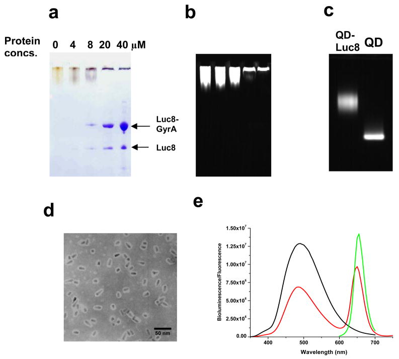
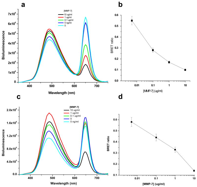
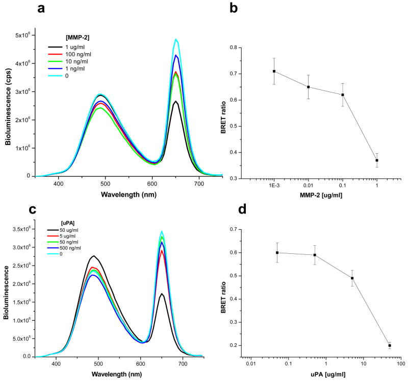
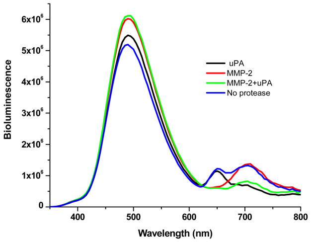
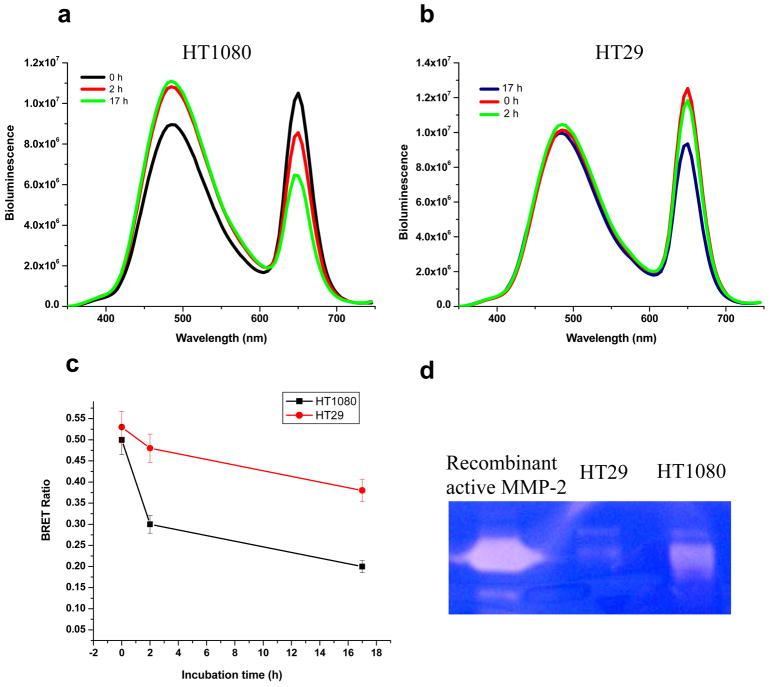
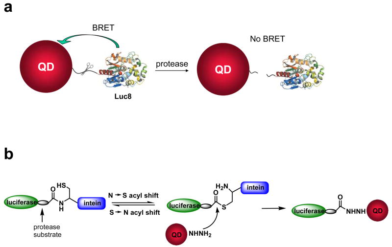
We report here a protease sensing nanoplatform based on semiconductor nanocrystals or quantum dots (QDs) and bioluminescence resonance energy transfer (QD-BRET) to detect the protease activity in complex biological samples. These nanosensors consist of bioluminescent proteins as the BRET donor, quantum dots as the BRET acceptor, and protease substrates sandwiched between the two as a sensing group. An intein-mediated conjugation strategy was developed for site-specific conjugation of proteins to QDs in preparing these QD nanosensors. In this traceless ligation, the intein itself is spliced out and excluded from the final conjugation product. With this method, we have synthesized a series of QD nanosensors for highly sensitive detection of an important class of protease matrix metalloproteinase (MMP) activity. We demonstrated that these nanosensors can detect the MMP activity in buffers and in mouse serum with the sensitivity to a few nanograms per milliliter and secreted proteases by tumor cells. The suitability of these nanosensors for a multiplex protease assay has also been shown.


Figures

Figure 1
Figure 1
Characterization of intein-mediated conjugation of Luc8 to QDs. The reactions were carried out with 4–40 μM of Luc8-GyrA fusion protein and 130 nM of QD655 hydrazide in 20 mM Tris buffer, pH 7.5 at room temperature for 2 h. a) SDS-PAGE gel analysis of conjugation reaction. b) Fluorescence image of the SDS-PAGE gel in a (excited at 302 nm). c) Fluorescence image of agarose gel of the unconjugated QDs and the conjugation reaction mixture. d) TEM micrograph of the prepared QD655-Luc8 conjugates (Note that the white halos around the particles represent proteins). e) Fluorescence emission spectrum of QD655 (λex = 480 nm; green), bioluminescent spectrum of Luc8 in the oxidation of coelenterazine (black), and bioluminescence emission spectrum of ligation product QD655-Luc8 in Tris buffer (red).
Figure 2
Figure 2
Detection of MMP-7 activity by nanosensor QD655-MMP-7-Luc8 in buffer (a, b) and in mouse serum (c, d). a) Representative emission spectra of QD655-MMP-7-Luc8 (0.3 μM) incubated with indicated MMP-7 concentrations for 1 h incubation in 20 mM Tris buffer (pH 7.5) at room temperature. The emission spectra were measured upon the addition of coelenterazine (1 μg). b) The BRET ratios in a) are plotted versus MMP-7 concentrations. c) Representative bioluminescence spectra of QD655-MMP-7-Luc8 (160 nM) incubated with indicated concentrations of MMP-7 in mouse serum for 1 h at room temperature. d) The BRET ratios in c) are plotted versus MMP-7 concentrations.
Figure 3
Figure 3
Detection of MMP-2 and uPA activity. a) Representative emission spectra of QD655-MMP-2-Luc8 (0.32 μM) incubated with indicated amount of MMP-2 in 20 mM Tris buffer (pH 7.5) for 1 h at room temperature. b) The BRET ratio in a) is plotted versus the MMP-2 concentrations. c) Representative emission spectra of QD655-uPA-Luc8 (0.32 μM) incubated with indicated amount of uPA in 20 mM Tris Buffer (pH 7.5) for 1 h at room temperature. d) The BRET ratio in c) is plotted versus the uPA concentration. All emission spectra were measured upon the addition of coelenterazine (1 μg).
Figure 4
Figure 4
Simultaneous detection of MMP-2 and uPA. A mixture of QD655-MMP-2-Luc8 (0.15 μM) and QD705-uPA-Luc8 (0.2 μM) were incubated with MMP-2 (1 μg/ml; red), uPA (10 μg/ml; black), MMP-2 (1 μg/ml) + uPA (10 μg/ml) (green), or no enzyme (blue) at room temperature for 1 h in 20 mM Tris buffer (pH 7.5).
Figure 5
Figure 5
Detection of MMP-2 activity in tumor cell culture media. a) Representative emission spectra of QD655-MMP-2-Luc8 (220 nM) incubated in HT1080 cell culture media for 0, 2, or 17 h. b) QD655-MMP-2-Luc8 (250 nM) was incubated in HT29 cell culture media for 0, 2, or 17 h. c) The plot of the BRET ratio changes in HT1080 and HT29 cell culture media over time. d) Gelatin zymograph assay of the MMP-2 activity in cell media culturing HT1080 and HT29; commercial recombinant active MMP-2 was used as the control.
Scheme 1
Scheme 1
Design and preparation of QD-BRET based nanosensors for MMP sensing. a) Schematic of the nanosensor comprising of a QD and luciferase proteins (Luc8) that are linked to the QD through an MMP peptide substrate. b) Intein-mediated site-specific conjugation of Luc8 fusion proteins to QDs.

References

    1. Fingleton B. Front Biosci. 2006;11:479–491. - PubMed
    1. Egeblad M, Werb Z. Nat Rev Cancer. 2002;2:161–174. - PubMed
    1. Brinckerhoff CE, Matrisian LM. Nat Rev Mol Cell Biol. 2002;3:207–214. - PubMed
    1. Bachmeier BE, Iancu CM, Jochum M, Nerlich AG. Expert Rev Anticancer Ther. 2005;5:149–163. - PubMed
    1. Overall CM, Kleifeld O. Nature Rev Cancer. 2006;6:227–239. - PubMed

Publication types

MeSH terms