. 2008 Nov 11;47(45):11704-10.
doi: 10.1021/bi8015982. Epub 2008 Oct 16.

Solution structure of Pyrococcus furiosus RPP21, a component of the archaeal RNase P holoenzyme, and interactions with its RPP29 protein partner

Affiliations

Solution structure of Pyrococcus furiosus RPP21, a component of the archaeal RNase P holoenzyme, and interactions with its RPP29 protein partner

Carlos D Amero et al. Biochemistry. .

Abstract

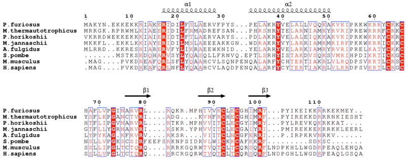
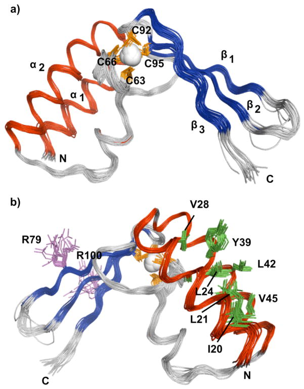
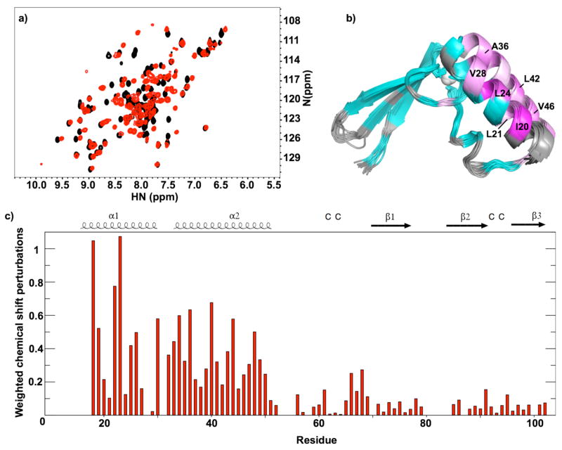
RNase P is the ubiquitous ribonucleoprotein metalloenzyme responsible for cleaving the 5'-leader sequence of precursor tRNAs during their maturation. While the RNA subunit is catalytically active on its own at high monovalent and divalent ion concentrations, four protein subunits are associated with archaeal RNase P activity in vivo: RPP21, RPP29, RPP30, and POP5. These proteins have been shown to function in pairs: RPP21-RPP29 and POP5-RPP30. We have determined the solution structure of RPP21 from the hyperthermophilic archaeon Pyrococcus furiosus ( Pfu) using conventional and paramagnetic NMR techniques. Pfu RPP21 in solution consists of an unstructured N-terminus, two alpha-helices, a zinc binding motif, and an unstructured C-terminus. Moreover, we have used chemical shift perturbations to characterize the interaction of RPP21 with RPP29. The data show that the primary contact with RPP29 is localized to the two helices of RPP21. This information represents a fundamental step toward understanding structure-function relationships of the archaeal RNase P holoenzyme.


Figures

Figure 1
Figure 1
Sequence alignment of RPP21 homologs from Archaea and Eukarya. Alignment was generated with CLUSTALW (47). Red lettering indicates a global similarity score of 0.7 while red boxes indicate invariant residues. Secondary structural elements observed in the NMR ensemble are indicated in cartoon format. The aligned sequences are from Pyrococcus furiousus, (NCBI code NP_579342); Methanothermobacter thermautotrophicus (NCBI code NP_276730); P. horikoshii (NCBI code NP_143456); Methanocaldococcus jannaschii (NCBI code NP_247957); Archaeoglobus fulgidus (NCBI code NP_068950); Schizosaccharomyces pombe (NCBI code NP_596472); Mus musculus (NCBI code NP_080584) and Homo sapiens (NCBI code NP_079115). Pfu RPP21 belongs to pfam04032. The figure was generated with ESPRIPT (48).
Figure 2
Figure 2
Two dimensional 1H-15N NMR spectra of Pfu RPP21. Backbone amide assignments as indicated. (a) Zinc-reconstituted RPP21. (b) Cobalt-reconstituted RPP21.
Figure 3
Figure 3
Solution structure of Pfu RPP21. (a) Ensemble of 20 lowest-energy structures superposed on the backbone heavy atoms of residues 19–82,88-105. (b) Rotated view showing the side-chains of some conserved residues.
Figure 4
Figure 4
Superposition of Pfu and Pho RPP21. Superposition of the core structural elements of a representative member from the NMR ensemble and the x-ray structure.
Figure 5
Figure 5
Binding of Pfu RPP21 to Pfu RPP29 as detected by NMR. a) Overlay of 15N HSQC spectra of Pfu RPP21 free (black) and Pfu RPP21 bound to Pfu RPP29 (red), illustrating the site-specific chemical shift perturbations that reveal the region of protein-protein interactions. b) Binding-induced chemical shift perturbations mapped onto the ribbon diagram of RPP21 structure, using a linear ramp from cyan (less than the mean perturbation) to magenta (normalized maximal perturbation). Residues for which the effect of RPP29 binding could not be determined are shown in light gray. c) Plot of weighted average chemical shift perturbations induced by protein-protein contacts.

Similar articles

Cited by

References

    1. Altman S, Baer MF, Bartkiewicz M, Gold H, Guerrier-Takada C, Kirsebom LA, Lumelsky N, Peck K. Catalysis by the RNA subunit of RNase P--a minireview. Gene. 1989;82:63–64. - PubMed
    1. Gopalan V, Altman S. In: Ribonuclease P: Structure and Catalysis, in The RNA World. Gesteland R, Cech T, Atkins J, editors. Cold Spring Harbor Laboratory Press; New York: 2006. only online at http://rna.cshl.edu)
    1. Evans D, Marquez SM, Pace NR. RNase P: interface of the RNA and protein worlds. Trends Biochem Sci. 2006;31:333–341. - PubMed
    1. Hall TA, Brown JW. Archaeal RNase P has multiple protein subunits homologous to eukaryotic nuclear RNase P proteins. RNA. 2002;8:296–306. - PMC - PubMed
    1. Tsai HY, Pulukkunat DK, Woznick WK, Gopalan V. Functional reconstitution and characterization of Pyrococcus furiosus RNase P. Proc Natl Acad Sci U S A. 2006;103:16147–16152. - PMC - PubMed

Publication types

MeSH terms