. 2008 Nov 11;47(45):11900-8.
doi: 10.1021/bi800981j. Epub 2008 Oct 16.

The efficacy of topoisomerase II-targeted anticancer agents reflects the persistence of drug-induced cleavage complexes in cells

Affiliations

The efficacy of topoisomerase II-targeted anticancer agents reflects the persistence of drug-induced cleavage complexes in cells

Omari J Bandele et al. Biochemistry. .

Abstract

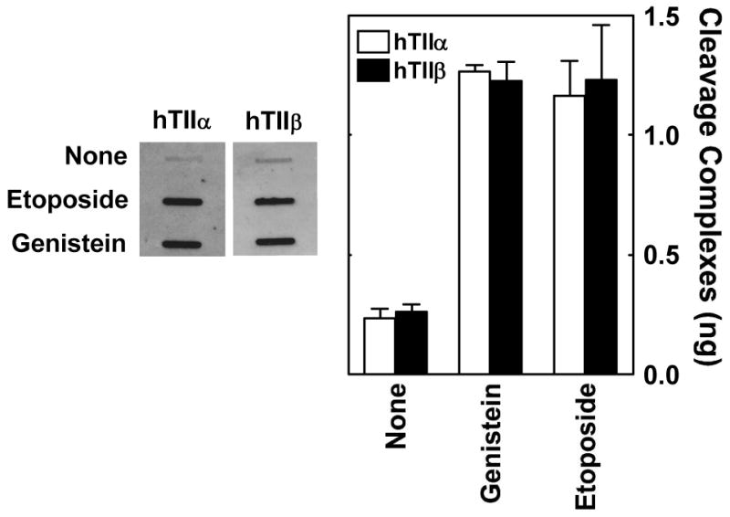
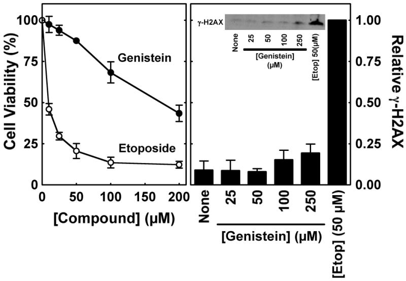
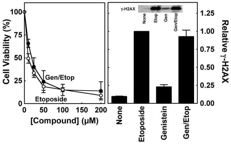
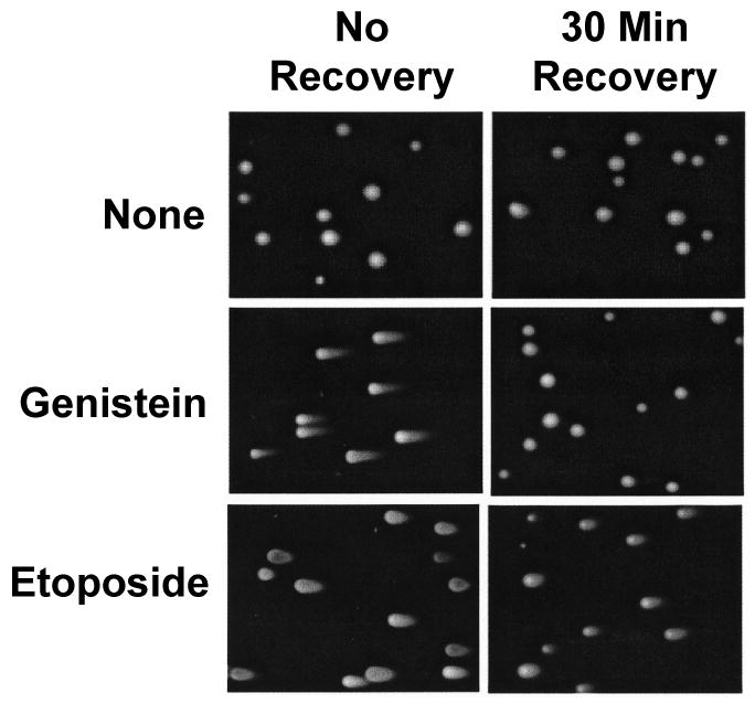
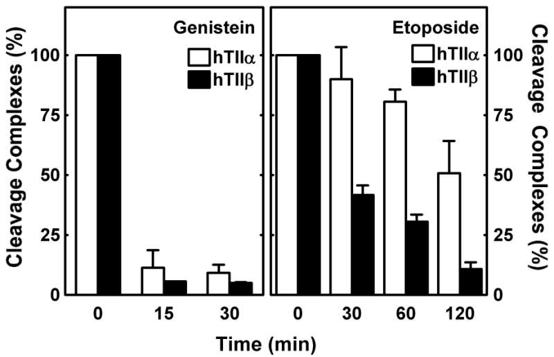
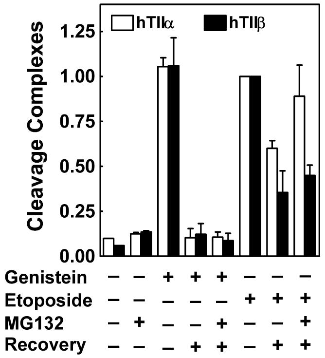
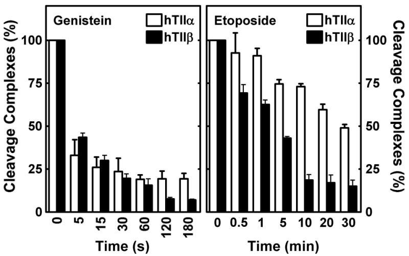
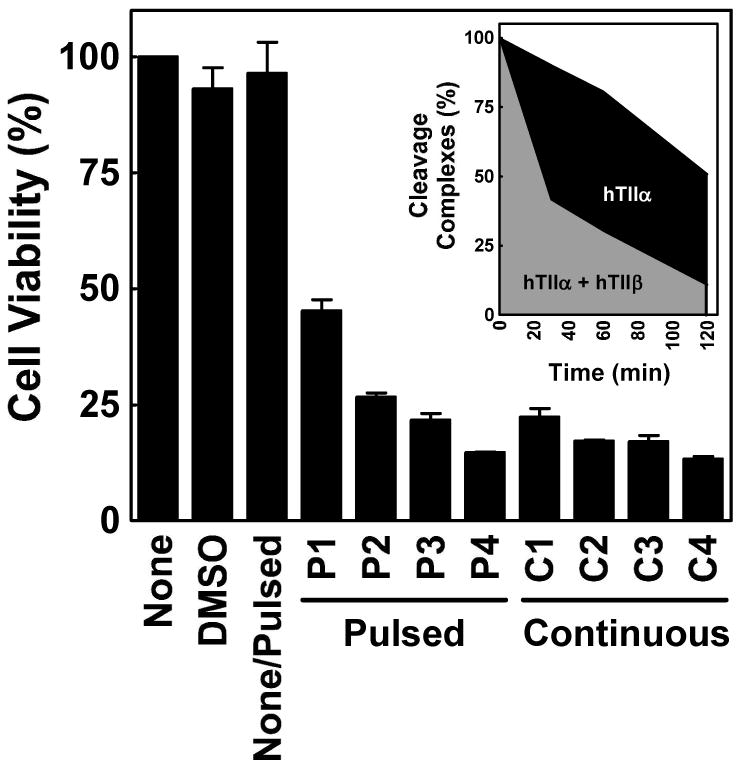
Genistein, a widely consumed bioflavonoid with chemopreventative properties in adults, and etoposide, a commonly prescribed anticancer drug, are well-characterized topoisomerase II poisons. Although both compounds display similar potencies against human topoisomerase IIalpha and IIbeta in vitro and induce comparable levels of DNA cleavage complexes in cultured human cells, their cytotoxic and genotoxic effects differ significantly. As determined by assays that monitored cell viability or the phosphorylation of histone H2AX, etoposide was much more toxic in CEM cells than genistein. Further studies that characterized the simultaneous treatment of cells with genistein and etoposide indicate that the differential actions of the two compounds are not related to the effects of genistein on cellular processes outside of its activity against topoisomerase II. Rather, they appear to result from a longer persistence of cleavage complexes induced by etoposide as compared to genistein. Parallel in vitro studies with purified type II enzymes led to similar conclusions regarding cleavage complex persistence. Isoform-specific differences were observed in vitro and in cells treated with etoposide. To this point, the t 1/2 of etoposide-induced DNA cleavage complexes formed with topoisomerase IIalpha in CEM cells was approximately 5 times longer than those formed with topoisomerase IIbeta. The cytotoxicity of etoposide following four treatment-recovery cycles was similar to that induced by continuous exposure to the drug over an equivalent time period. Taken together, these findings suggest that it may be possible to preferentially target topoisomerase IIalpha with etoposide by employing a schedule that utilizes pulsed drug treatment-recovery cycles.


Figures

FIGURE 1
FIGURE 1
Ability of genistein and etoposide to enhance DNA cleavage mediated by topoisomerase IIα and β in cultured human CEM cells. The ICE bioassay was used to monitor levels of isoform-specific cleavage complexes in cells treated with genistein or etoposide. DNA from cell cultures treated for 1 h in the absence of compound (None) or in the presence of 50 μM genistein or etoposide was blotted onto a nitrocellulose membrane. Immunoblots were probed with polyclonal antibodies directed against either human topoisomerase IIα or β. A representative immunoblot is shown (left). The bar graph shows data for topoisomerase IIα (hTIIα; open bars) and β (hTIIβ; closed bars). Levels of covalently bound topoisomerase II are based on standards of purified human type II topoisomerases. Error bars represent the standard deviation for three independent experiments. Data were adapted from Ref. .
FIGURE 2
FIGURE 2
Cytotoxic and genotoxic effects of genistein and etoposide in cultured human CEM cells. Left Panel: The viability of cells in the presence of genistein or etoposide was monitored using the CCK-8 cell proliferation assay. Data are shown for cells treated with 0–200 μM genistein (closed circles) or etoposide (open circles) for 8 h. Cell viability was set to 100% in the absence of compounds. Right Panel: Phosphorylation of histone H2AX (i.e., γ-H2AX) was monitored as a marker for permanent DNA damage in CEM cells. Data are shown for cells treated in the absence of compound (None) or in the presence of 0–250 μM genistein or 50 μM etoposide for 1 h. The inset shows a representative immunoblot that was probed with a monoclonal antibody directed against γ-H2AX (Ser 139). Levels of permanent DNA damage are relative to those in the presence of 50 μM etoposide (set to 1.0). Error bars represent the standard deviation for three independent experiments.
FIGURE 3
FIGURE 3
Effects of genistein on the cytotoxicity and genotoxicity of etoposide in cultured human CEM cells. Left Panel: The CCK-8 cell proliferation assay was used to monitor the viability of cells incubated with 0–200 μM etoposide alone (open circles) or 50 μM genistein for 2 h prior to and during a 6 h exposure to 0–200 μM etoposide (closed circles; Gen/Etop). Cell viability was set to 100% in the absence of compounds. Right Panel: Phosphorylation of histone H2AX (i.e., γ-H2AX) was monitored as a marker for permanent DNA damage to examine the effect of genistein on the genotoxicity of etoposide in CEM cells. Data are shown for cells treated in the absence of compound (None), in the presence of 50 μM etoposide alone for 1 h (Etoposide) or 50 μM genistein alone for 3 h (Genistein), or in the presence of 50 μM genistein for 2 h prior to and during a 1 h exposure to 50 μM etoposide (Gen/Etop). The inset shows a representative immunoblot that was probed with a monoclonal antibody directed against γ-H2AX (Ser 139). Levels of γ-H2AX are relative to those in the presence of etoposide (set to 1.0). Error bars represent the standard deviation for three independent experiments.
FIGURE 4
FIGURE 4
Effects of genistein and etoposide on the persistence of DNA cleavage complexes mediated by topoisomerase II in cultured human CEM cells. The neutral comet assay was utilized to monitor the levels of double-stranded DNA breaks in treated cells. Left panels show representative cells treated in the absence of compounds (None) or in the presence of 50 μM genistein or etoposide for 1 h and lysed immediately following treatment (i.e., No Recovery) Right panels show representative cells from parallel experiments in which cultures were transferred to drug-free medium for 30 min prior to lysis (i.e., 30 Min Recovery). The length of the “comet tail” of each cell is proportional to the level of double-stranded DNA breaks. Data are representative of at least three independent experiments. At least ten fields similar to those shown were observed for each set of experimental conditions.
FIGURE 5
FIGURE 5
Effects of genistein and etoposide on the persistence of topoisomerase IIα- and β-DNA cleavage complexes in cultured human CEM cells. The ICE bioassay was utilized to monitor levels of isoform-specific DNA cleavage complexes induced by genistein (left panel) or etoposide (right panel). Cells were treated with 50 μM compounds for 1 h and transferred to drug-free medium for the indicated times. Data for topoisomerase IIα- (hTIIα; open bars) and topoisomerase IIβ-DNA cleavage complexes (hTIIβ; closed bars) are shown. Relative levels of complexes prior to recovery were set to 100%. Error bars represent the standard deviation for three independent experiments.
FIGURE 6
FIGURE 6
Role of the 26S proteasome in the degradation of topoisomerase IIα– and β–DNA cleavage complexes induced by genistein and etoposide. The ICE bioassay was utilized to monitor levels of isoform-specific DNA cleavage complexes. CEM cells were treated in the absence of compound or in the presence of 2 μM MG132 (a proteasome inhibitor), 50 μM genistein, or 50 μM etoposide for 1 h. In some cases, cells were transferred to drug-free medium for a 30 min recovery period following treatment. When experiments were carried out in presence of MG132 (2 μM), the proteasome inhibitor was included in cell cultures for 30 min prior to the addition of genistein or etoposide and also was present during the recovery period. Data for cleavage complexes mediated by topoisomerase IIα (hTIIα; open bars) or β (hTIIβ; closed bars) are shown. Levels of cleavage complexes prior to recovery were set to 100%. Error bars represent the standard deviation for three independent experiments.
FIGURE 7
FIGURE 7
Effects of genistein and etoposide on the persistence of topoisomerase IIα- and β-DNA cleavage complexes in vitro. DNA cleavage assays were performed using purified human type II enzymes. Reactions were initiated in the presence of 50 μM genistein (left panel) or etoposide (right panel). After cleavage equilibrium was established (6 min), reaction mixtures were diluted 10–fold with DNA cleavage buffer, and levels of double-stranded DNA breaks were monitored over time. Data for DNA cleavage mediated by topoisomerase IIα (hTIIα; open bars) or β (hTIIβ; closed bars) are shown. Relative levels of DNA cleavage prior to dilution were set to 100%. Error bars represent the standard deviation for three independent experiments.
FIGURE 8
FIGURE 8
Cytotoxicity of etoposide using a pulsed treatment schedule in cultured human CEM cells. The CCK-8 proliferation assay was utilized to compare the viability of cells treated with a pulsed schedule (4 cycles of treatment with etoposide for 15 min followed by a 2 h recovery period in drug-free medium) vs. a continuous exposure to 50 μM etoposide for an equivalent period of time (9 h total). Control data are shown for cells treated in the absence of drug (None), in the continuous presence of drug diluent (DMSO), or in the absence of drug but subjected to the physical manipulation of the pulsed schedule (None/Pulsed). The viability of cells is shown following each cycle of the pulsed schedule (Cycle P1-4) and for samples that were removed at equivalent times (2.25 h intervals) for cultures that were continuously exposed to etoposide (Cycle C1-4). Cell viability was set to 100% in the absence of drug. Error bars represent the standard deviation for three independent experiments. The inset shows the area under the curve for levels of etoposide-induced DNA cleavage complexes mediated by topoisomerase IIα (black + gray filled areas) and β (gray filled area) for one pulse-recovery cycle, as monitored by the ICE bioassay.

References

    1. Berger JM, Gamblin SJ, Harrison SC, Wang JC. Structure and mechanism of DNA topoisomerase II. Nature. 1996;379:225–232. - PubMed
    1. Nitiss JL. Investigating the biological functions of DNA topoisomerases in eukaryotic cells. Biochim Biophys Acta. 1998;1400:63–81. - PubMed
    1. Fortune JM, Osheroff N. Topoisomerase II as a target for anticancer drugs: When enzymes stop being nice. Prog Nucleic Acid Res Mol Biol. 2000;64:221–253. - PubMed
    1. Champoux JJ. DNA topisomerases: Structure, function, and mechanism. Annu Rev Biochem. 2001;70:369–413. - PubMed
    1. Wang JC. Cellular roles of DNA topoisomerases: A molecular perspective. Nat Rev Mol Cell Biol. 2002;3:430–440. - PubMed

Publication types